6WVZ

Crystal structure of anti-MET Fab arm of amivantamab in complex with human MET


Experimental Data Snapshot

  • Method: X-RAY DIFFRACTION
  • Resolution: 3.10 Å
  • R-Value Free: 0.235 
  • R-Value Work: 0.190 
  • R-Value Observed: 0.192 

wwPDB Validation   3D Report Full Report


Ligand Structure Quality Assessment 


This is version 1.2 of the entry. See complete history


Literature

Discovery of amivantamab (JNJ-61186372), a bispecific antibody targeting EGFR and MET.

Neijssen, J.Cardoso, R.M.F.Chevalier, K.M.Wiegman, L.Valerius, T.Anderson, G.M.Moores, S.L.Schuurman, J.Parren, P.W.H.I.Strohl, W.R.Chiu, M.L.

(2021) J Biol Chem 296: 100641-100641

  • DOI: https://doi.org/10.1016/j.jbc.2021.100641
  • Primary Citation of Related Structures:  
    6WVZ

  • PubMed Abstract: 

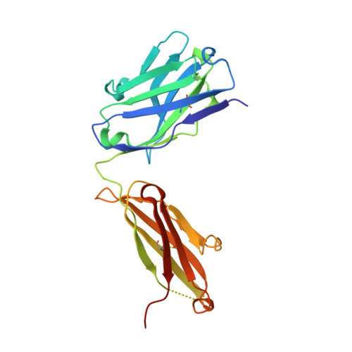
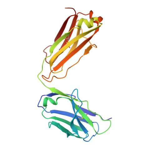
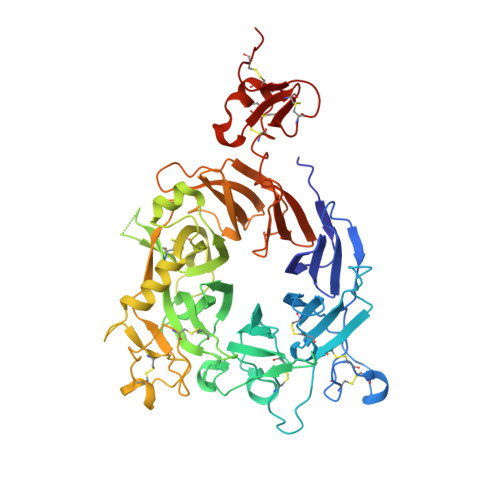
    A bispecific antibody (BsAb) targeting the epidermal growth factor receptor (EGFR) and mesenchymal-epithelial transition factor (MET) pathways represents a novel approach to overcome resistance to targeted therapies in patients with non-small cell lung cancer. In this study, we sequentially screened a panel of BsAbs in a combinatorial approach to select the optimal bispecific molecule. The BsAbs were derived from different EGFR and MET parental monoclonal antibodies. Initially, molecules were screened for EGFR and MET binding on tumor cell lines and lack of agonistic activity toward MET. Hits were identified and further screened based on their potential to induce untoward cell proliferation and cross-phosphorylation of EGFR by MET via receptor colocalization in the absence of ligand. After the final step, we selected the EGFR and MET arms for the lead BsAb and added low fucose Fc engineering to generate amivantamab (JNJ-61186372). The crystal structure of the anti-MET Fab of amivantamab bound to MET was solved, and the interaction between the two molecules in atomic details was elucidated. Amivantamab antagonized the hepatocyte growth factor (HGF)-induced signaling by binding to MET Sema domain and thereby blocking HGF β-chain-Sema engagement. The amivantamab EGFR epitope was mapped to EGFR domain III and residues K443, K465, I467, and S468. Furthermore, amivantamab showed superior antitumor activity over small molecule EGFR and MET inhibitors in the HCC827-HGF in vivo model. Based on its unique mode of action, amivantamab may provide benefit to patients with malignancies associated with aberrant EGFR and MET signaling.


  • Organizational Affiliation

    Genmab, Utrecht, The Netherlands.


Macromolecules
Find similar proteins by:  (by identity cutoff)  |  3D Structure
Entity ID: 1
MoleculeChains Sequence LengthOrganismDetailsImage
Heavy Chain of anti-MET Fab of amivantamabA [auth H]228Homo sapiensMutation(s): 0 
Entity Groups  
Sequence Clusters30% Identity50% Identity70% Identity90% Identity95% Identity100% Identity
Sequence Annotations
Expand
  • Reference Sequence
Find similar proteins by:  (by identity cutoff)  |  3D Structure
Entity ID: 2
MoleculeChains Sequence LengthOrganismDetailsImage
Light Chain of anti-MET Fab of amivantamabB [auth L]214Homo sapiensMutation(s): 0 
Entity Groups  
Sequence Clusters30% Identity50% Identity70% Identity90% Identity95% Identity100% Identity
Sequence Annotations
Expand
  • Reference Sequence
Find similar proteins by:  (by identity cutoff)  |  3D Structure
Entity ID: 3
MoleculeChains Sequence LengthOrganismDetailsImage
Hepatocyte growth factor receptorC [auth M]534Homo sapiensMutation(s): 0 
Gene Names: MET
EC: 2.7.10.1
UniProt & NIH Common Fund Data Resources
Find proteins for P08581 (Homo sapiens)
Explore P08581 
Go to UniProtKB:  P08581
PHAROS:  P08581
GTEx:  ENSG00000105976 
Entity Groups  
Sequence Clusters30% Identity50% Identity70% Identity90% Identity95% Identity100% Identity
UniProt GroupP08581
Sequence Annotations
Expand
  • Reference Sequence
Oligosaccharides

Help

Entity ID: 4
MoleculeChains Length2D Diagram Glycosylation3D Interactions
2-acetamido-2-deoxy-beta-D-glucopyranose-(1-4)-2-acetamido-2-deoxy-beta-D-glucopyranoseD [auth A],
F [auth C]
2N-Glycosylation
Glycosylation Resources
GlyTouCan:  G42666HT
GlyCosmos:  G42666HT
GlyGen:  G42666HT
Entity ID: 5
MoleculeChains Length2D Diagram Glycosylation3D Interactions
2-acetamido-2-deoxy-beta-D-glucopyranose-(1-4)-[alpha-L-fucopyranose-(1-6)]2-acetamido-2-deoxy-beta-D-glucopyranoseE [auth B],
G [auth D]
3N-Glycosylation
Glycosylation Resources
GlyTouCan:  G21290RB
GlyCosmos:  G21290RB
GlyGen:  G21290RB
Small Molecules
Ligands 2 Unique
IDChains Name / Formula / InChI Key2D Diagram3D Interactions
NAG
Query on NAG

Download Ideal Coordinates CCD File 
H [auth M]2-acetamido-2-deoxy-beta-D-glucopyranose
C8 H15 N O6
OVRNDRQMDRJTHS-FMDGEEDCSA-N
FMT
Query on FMT

Download Ideal Coordinates CCD File 
I [auth M]FORMIC ACID
C H2 O2
BDAGIHXWWSANSR-UHFFFAOYSA-N
Experimental Data & Validation

Experimental Data

  • Method: X-RAY DIFFRACTION
  • Resolution: 3.10 Å
  • R-Value Free: 0.235 
  • R-Value Work: 0.190 
  • R-Value Observed: 0.192 
  • Space Group: P 43 21 2
Unit Cell:
Length ( Å )Angle ( ˚ )
a = 86.83α = 90
b = 86.83β = 90
c = 457.099γ = 90
Software Package:
Software NamePurpose
HKL-2000data reduction
HKL-2000data scaling
PHASERphasing
PHENIXrefinement
PDB_EXTRACTdata extraction

Structure Validation

View Full Validation Report


Ligand Structure Quality Assessment 


Entry History 

Deposition Data

Revision History  (Full details and data files)

  • Version 1.0: 2021-04-28
    Type: Initial release
  • Version 1.1: 2021-07-21
    Changes: Database references
  • Version 1.2: 2023-10-18
    Changes: Data collection, Database references, Refinement description