Covalent Inactivation of Mycobacterium tuberculosis Isocitrate Lyase by cis -2,3-Epoxy-Succinic Acid.
Pham, T.V., Mellott, D.M., Moghadamchargari, Z., Chen, K., Krieger, I., Laganowsky, A., Sacchettini, J.C., Meek, T.D.(2021) ACS Chem Biol 16: 463-470
- PubMed: 33688722 Search on PubMed
- DOI: https://doi.org/10.1021/acschembio.0c00740
- Primary Citation Related Structures:
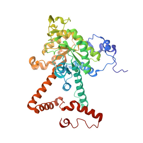
6VB9, 6WSI - PubMed Abstract:
The isocitrate lyases (ICL1/2) are essential enzymes of Mycobacterium tuberculosis ( Mtb ), the causative agent of tuberculosis. At present, no ICL1/2 inhibitors have progressed to clinical evaluation, despite extensive drug discovery efforts. Herein, we surveyed succinate analogs against ICL1 and found that dicarboxylic acids constrained in their synperiplanar conformations, such as maleic acid, comprise uncompetitive inhibitors of ICL1 and inhibit more potently than their trans -isomers. From this, we identified cis -2,3 epoxysuccinic acid ( cis -EpS) as a selective, irreversible covalent inactivator of Mtb ICL1 ( k inact / K inact = (5.0 ± 1.4) × 10 4 M -1 s -1 ; K inact = 200 ± 50 nM), the most potent inactivator of ICL1 yet characterized. Crystallographic and mass spectrometric analysis demonstrated that Cys 191 of ICL1 was S-malylated by cis -EpS, and a crystallographic "snapshot" of inactivation lent insight into the chemical mechanism of this inactivation. Proteomic analysis of E. coli lysates showed that cis -EpS selectively labeled plasmid-expressed Mtb ICL1. Consistently, cis -EpS, but not its trans -isomer, inhibited the growth of Mtb under conditions in which ICL function is essential. These findings encourage the development of analogs of cis -2,3-epoxysuccinate as antituberculosis agents.
- Department of Biochemistry and Biophysics, Texas A&M University, College Station, Texas 77843, United States.
Organizational Affiliation: