Structural analysis of the catalytic domain of Artemis endonuclease/SNM1C reveals distinct structural features.
Karim, M.F., Liu, S., Laciak, A.R., Volk, L., Koszelak-Rosenblum, M., Lieber, M.R., Wu, M., Curtis, R., Huang, N.N., Carr, G., Zhu, G.(2020) J Biological Chem 295: 12368-12377
- PubMed: 32576658 Search on PubMedSearch on PubMed Central
- DOI: https://doi.org/10.1074/jbc.RA120.014136
- Primary Citation Related Structures:
6WNL, 6WO0 - PubMed Abstract:
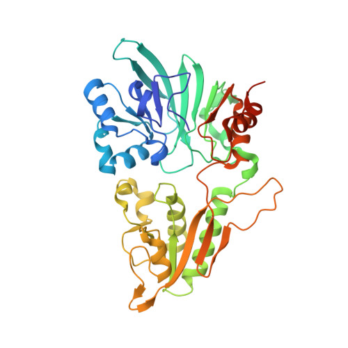
The endonuclease Artemis is responsible for opening DNA hairpins during V(D)J recombination and for processing a subset of pathological DNA double-strand breaks. Artemis is an attractive target for the development of therapeutics to manage various B cell and T cell tumors, because failure to open DNA hairpins and accumulation of chromosomal breaks may reduce the proliferation and viability of pre-T and pre-B cell derivatives. However, structure-based drug discovery of specific Artemis inhibitors has been hampered by a lack of crystal structures. Here, we report the structure of the catalytic domain of recombinant human Artemis. The catalytic domain displayed a polypeptide fold similar overall to those of other members in the DNA cross-link repair gene SNM1 family and in mRNA 3'-end-processing endonuclease CPSF-73, containing metallo-β-lactamase and β-CASP domains and a cluster of conserved histidine and aspartate residues capable of binding two metal atoms in the catalytic site. As in SNM1A, only one zinc ion was located in the Artemis active site. However, Artemis displayed several unique features. Unlike in other members of this enzyme class, a second zinc ion was present in the β-CASP domain that leads to structural reorientation of the putative DNA-binding surface and extends the substrate-binding pocket to a new pocket, pocket III. Moreover, the substrate-binding surface exhibited a dominant and extensive positive charge distribution compared with that in the structures of SNM1A and SNM1B, presumably because of the structurally distinct DNA substrate of Artemis. The structural features identified here may provide opportunities for designing selective Artemis inhibitors.
- Discovery Biology, Albany Molecular Research Inc., Buffalo, New York, USA.
Organizational Affiliation: