6WN6

Crystal structure of 3-keto-D-glucoside 4-epimerase, YcjR, from E. coli, apo form


Experimental Data Snapshot

  • Method: X-RAY DIFFRACTION
  • Resolution: 1.86 Å
  • R-Value Free: 0.199 
  • R-Value Work: 0.175 
  • R-Value Observed: 0.176 

wwPDB Validation   3D Report Full Report


Ligand Structure Quality Assessment 


This is version 1.2 of the entry. See complete history


Literature

Structure and Reaction Mechanism of YcjR, an Epimerase That Facilitates the Interconversion of d-Gulosides to d-Glucosides inEscherichia coli.

Mabanglo, M.F.Huddleston, J.P.Mukherjee, K.Taylor, Z.W.Raushel, F.M.

(2020) Biochemistry 59: 2069-2077

  • DOI: https://doi.org/10.1021/acs.biochem.0c00334
  • Primary Citation of Related Structures:  
    6WN6

  • PubMed Abstract: 

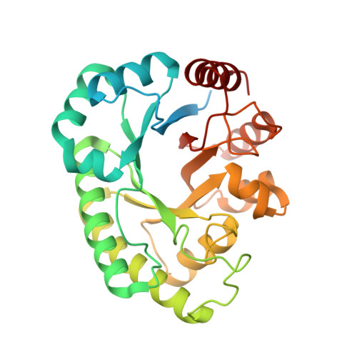
    YcjR from Escherichia coli K-12 MG1655 catalyzes the manganese-dependent reversible epimerization of 3-keto-α-d-gulosides to the corresponding 3-keto-α-d-glucosides as a part of a proposed catabolic pathway for the transformation of d-gulosides to d-glucosides. The three-dimensional structure of the manganese-bound enzyme was determined by X-ray crystallography. The divalent manganese ion is coordinated to the enzyme by ligation to Glu-146, Asp-179, His-205, and Glu-240. When either of the two active site glutamate residues is mutated to glutamine, the enzyme loses all catalytic activity for the epimerization of α-methyl-3-keto-d-glucoside at C4. However, the E240Q mutant can catalyze hydrogen-deuterium exchange of the proton at C4 of α-methyl-3-keto-d-glucoside in solvent D 2 O. The E146Q mutant does not catalyze this exchange reaction. These results indicate that YcjR catalyzes the isomerization of 3-keto-d-glucosides via proton abstraction at C4 by Glu-146 to form a cis -enediolate intermediate that is subsequently protonated on the opposite face by Glu-240 to generate the corresponding 3-keto-d-guloside. This conclusion is supported by docking of the cis -enediolate intermediate into the active site of YcjR based on the known binding orientation of d-fructose and d-psicose in the active site of d-psicose-3-epimerase.


  • Organizational Affiliation

    Department of Chemistry, Texas A&M University, College Station, Texas 77843, United States.


Macromolecules
Find similar proteins by:  (by identity cutoff)  |  3D Structure
Entity ID: 1
MoleculeChains Sequence LengthOrganismDetailsImage
3-keto-D-glucoside 4-epimerase
A, B, C, D
314Escherichia coliMutation(s): 0 
Gene Names: ycjRPGD_01932
UniProt
Find proteins for P76044 (Escherichia coli (strain K12))
Explore P76044 
Go to UniProtKB:  P76044
Entity Groups  
Sequence Clusters30% Identity50% Identity70% Identity90% Identity95% Identity100% Identity
UniProt GroupP76044
Sequence Annotations
Expand
  • Reference Sequence
Small Molecules
Ligands 2 Unique
IDChains Name / Formula / InChI Key2D Diagram3D Interactions
EDO (Subject of Investigation/LOI)
Query on EDO

Download Ideal Coordinates CCD File 
AA [auth B]
AB [auth C]
BA [auth B]
BB [auth C]
CA [auth B]
AA [auth B],
AB [auth C],
BA [auth B],
BB [auth C],
CA [auth B],
CB [auth C],
DA [auth B],
EA [auth B],
EB [auth D],
F [auth A],
FA [auth B],
FB [auth D],
G [auth A],
GA [auth B],
GB [auth D],
H [auth A],
HA [auth B],
HB [auth D],
I [auth A],
IA [auth B],
IB [auth D],
J [auth A],
JA [auth B],
JB [auth D],
K [auth A],
KB [auth D],
L [auth A],
LA [auth C],
LB [auth D],
M [auth A],
MA [auth C],
MB [auth D],
N [auth A],
NA [auth C],
NB [auth D],
O [auth A],
OA [auth C],
OB [auth D],
P [auth A],
PA [auth C],
PB [auth D],
Q [auth A],
QA [auth C],
QB [auth D],
R [auth A],
RA [auth C],
RB [auth D],
S [auth A],
SA [auth C],
SB [auth D],
T [auth A],
TA [auth C],
TB [auth D],
UA [auth C],
V [auth B],
VA [auth C],
W [auth B],
WA [auth C],
X [auth B],
XA [auth C],
Y [auth B],
YA [auth C],
Z [auth B],
ZA [auth C]
1,2-ETHANEDIOL
C2 H6 O2
LYCAIKOWRPUZTN-UHFFFAOYSA-N
MN
Query on MN

Download Ideal Coordinates CCD File 
DB [auth D],
E [auth A],
KA [auth C],
U [auth B]
MANGANESE (II) ION
Mn
WAEMQWOKJMHJLA-UHFFFAOYSA-N
Experimental Data & Validation

Experimental Data

  • Method: X-RAY DIFFRACTION
  • Resolution: 1.86 Å
  • R-Value Free: 0.199 
  • R-Value Work: 0.175 
  • R-Value Observed: 0.176 
  • Space Group: P 41 21 2
Unit Cell:
Length ( Å )Angle ( ˚ )
a = 116.576α = 90
b = 116.576β = 90
c = 247.725γ = 90
Software Package:
Software NamePurpose
HKL-2000data scaling
PHENIXrefinement
PDB_EXTRACTdata extraction
HKL-2000data reduction
PHASERphasing

Structure Validation

View Full Validation Report


Ligand Structure Quality Assessment 


Entry History & Funding Information

Deposition Data


Funding OrganizationLocationGrant Number
Robert A. Welch FoundationUnited StatesA-840

Revision History  (Full details and data files)

  • Version 1.0: 2020-06-10
    Type: Initial release
  • Version 1.1: 2020-06-17
    Changes: Database references
  • Version 1.2: 2024-03-06
    Changes: Data collection, Database references