6W5B

N124D Deamidation Mutant of Human gammaD-Crystallin


Experimental Data Snapshot

  • Method: X-RAY DIFFRACTION
  • Resolution: 1.15 Å
  • R-Value Free: 0.172 
  • R-Value Work: 0.151 
  • R-Value Observed: 0.152 

wwPDB Validation   3D Report Full Report


This is version 1.2 of the entry. See complete history


Literature

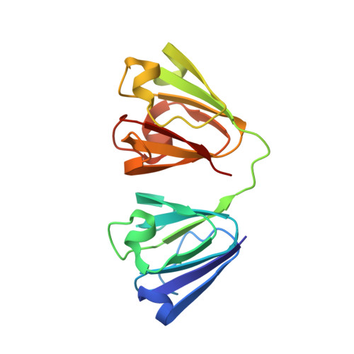
Assessing the Structures and Interactions of gamma D-Crystallin Deamidation Variants.

Guseman, A.J.Whitley, M.J.Gonzalez, J.J.Rathi, N.Ambarian, M.Gronenborn, A.M.

(2021) Structure 29: 284-291.e3

  • DOI: https://doi.org/10.1016/j.str.2020.11.006
  • Primary Citation of Related Structures:  
    6W5B, 6WCY

  • PubMed Abstract: 

    Cataracts involve the deposition of the crystallin proteins in the vertebrate eye lens, causing opacification and blindness. They are associated with either genetic mutation or protein damage that accumulates over the lifetime of the organism. Deamidation of Asn residues in several different crystallins has been observed and is frequently invoked as a cause of cataract. Here, we investigated the properties of Asp variants, deamidation products of γD-crystallin, by solution NMR, X-ray crystallography, and other biophysical techniques. No substantive structural or stability changes were noted for all seven Asn to Asp γD-crystallins. Importantly, no changes in diffusion interaction behavior could be detected. Our combined experimental results demonstrate that introduction of single Asp residues on the surface of γD-crystallin by deamidation is unlikely to be the driver of cataract formation in the eye lens.


  • Organizational Affiliation

    Department of Structural Biology, University of Pittsburgh School of Medicine, 3501 Fifth Avenue, Pittsburgh, PA 15261, USA.


Macromolecules
Find similar proteins by:  (by identity cutoff)  |  3D Structure
Entity ID: 1
MoleculeChains Sequence LengthOrganismDetailsImage
Gamma-crystallin D173Homo sapiensMutation(s): 1 
Gene Names: CRYGDCRYG4
UniProt & NIH Common Fund Data Resources
Find proteins for P07320 (Homo sapiens)
Explore P07320 
Go to UniProtKB:  P07320
PHAROS:  P07320
GTEx:  ENSG00000118231 
Entity Groups  
Sequence Clusters30% Identity50% Identity70% Identity90% Identity95% Identity100% Identity
UniProt GroupP07320
Sequence Annotations
Expand
  • Reference Sequence
Experimental Data & Validation

Experimental Data

  • Method: X-RAY DIFFRACTION
  • Resolution: 1.15 Å
  • R-Value Free: 0.172 
  • R-Value Work: 0.151 
  • R-Value Observed: 0.152 
  • Space Group: P 21 21 21
Unit Cell:
Length ( Å )Angle ( ˚ )
a = 33.649α = 90
b = 51.493β = 90
c = 93.794γ = 90
Software Package:
Software NamePurpose
PHENIXrefinement
XDSdata reduction
XDSdata scaling
PHASERphasing

Structure Validation

View Full Validation Report


Entry History & Funding Information

Deposition Data


Funding OrganizationLocationGrant Number
National Institutes of Health/National Eye Institute (NIH/NEI)United StatesEY030057

Revision History  (Full details and data files)

  • Version 1.0: 2021-01-20
    Type: Initial release
  • Version 1.1: 2021-03-17
    Changes: Database references
  • Version 1.2: 2023-10-18
    Changes: Data collection, Database references, Refinement description