6W4L

The crystal structure of a single chain H2B-H2A histone chimera from Xenopus laevis


Experimental Data Snapshot

  • Method: X-RAY DIFFRACTION
  • Resolution: 1.31 Å
  • R-Value Free: 0.206 
  • R-Value Work: 0.170 
  • R-Value Observed: 0.171 

wwPDB Validation   3D Report Full Report


This is version 1.2 of the entry. See complete history


Literature

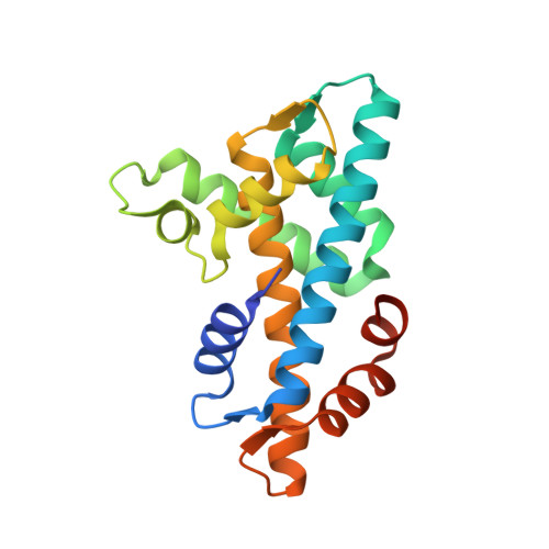
Structure of a single-chain H2A/H2B dimer.

Warren, C.Bonanno, J.B.Almo, S.C.Shechter, D.

(2020) Acta Crystallogr F Struct Biol Commun 76: 194-198

  • DOI: https://doi.org/10.1107/S2053230X20004604
  • Primary Citation of Related Structures:  
    6W4L

  • PubMed Abstract: 

    Chromatin is the complex assembly of nucleic acids and proteins that makes up the physiological form of the eukaryotic genome. The nucleosome is the fundamental repeating unit of chromatin, and is composed of ∼147 bp of DNA wrapped around a histone octamer formed by two copies of each core histone: H2A, H2B, H3 and H4. Prior to nucleosome assembly, and during histone eviction, histones are typically assembled into soluble H2A/H2B dimers and H3/H4 dimers and tetramers. A multitude of factors interact with soluble histone dimers and tetramers, including chaperones, importins, histone-modifying enzymes and chromatin-remodeling enzymes. It is still unclear how many of these proteins recognize soluble histones; therefore, there is a need for new structural tools to study non-nucleosomal histones. Here, a single-chain, tailless Xenopus H2A/H2B dimer was created by directly fusing the C-terminus of H2B to the N-terminus of H2A. It is shown that this construct (termed scH2BH2A) is readily expressed in bacteria and can be purified under non-denaturing conditions. A 1.31 Å resolution crystal structure of scH2BH2A shows that it adopts a conformation that is nearly identical to that of nucleosomal H2A/H2B. This new tool is likely to facilitate future structural studies of many H2A/H2B-interacting proteins.


  • Organizational Affiliation

    Department of Biochemistry, Albert Einstein College of Medicine, 1300 Morris Park Avenue, Bronx, NY 10461, USA.


Macromolecules
Find similar proteins by:  (by identity cutoff)  |  3D Structure
Entity ID: 1
MoleculeChains Sequence LengthOrganismDetailsImage
Histone H2B 1.1,Histone H2A type 1186Xenopus laevisMutation(s): 0 
UniProt
Find proteins for P06897 (Xenopus laevis)
Explore P06897 
Go to UniProtKB:  P06897
Find proteins for P02281 (Xenopus laevis)
Explore P02281 
Go to UniProtKB:  P02281
Entity Groups  
Sequence Clusters30% Identity50% Identity70% Identity90% Identity95% Identity100% Identity
UniProt GroupsP02281P06897
Sequence Annotations
Expand
  • Reference Sequence
Small Molecules
Ligands 1 Unique
IDChains Name / Formula / InChI Key2D Diagram3D Interactions
PPV
Query on PPV

Download Ideal Coordinates CCD File 
B [auth A]PYROPHOSPHATE
H4 O7 P2
XPPKVPWEQAFLFU-UHFFFAOYSA-N
Experimental Data & Validation

Experimental Data

  • Method: X-RAY DIFFRACTION
  • Resolution: 1.31 Å
  • R-Value Free: 0.206 
  • R-Value Work: 0.170 
  • R-Value Observed: 0.171 
  • Space Group: C 1 2 1
Unit Cell:
Length ( Å )Angle ( ˚ )
a = 61.793α = 90
b = 44.679β = 117.27
c = 66.45γ = 90
Software Package:
Software NamePurpose
Aimlessdata scaling
REFMACrefinement
PDB_EXTRACTdata extraction
XDSdata reduction
MOLREPphasing
Cootmodel building

Structure Validation

View Full Validation Report


Entry History & Funding Information

Deposition Data


Funding OrganizationLocationGrant Number
National Institutes of Health/National Institute of General Medical Sciences (NIH/NIGMS)United StatesR01GM135614

Revision History  (Full details and data files)

  • Version 1.0: 2020-05-06
    Type: Initial release
  • Version 1.1: 2020-05-13
    Changes: Database references
  • Version 1.2: 2023-10-18
    Changes: Data collection, Database references, Refinement description