6W4I

APE1 Y269A product complex with abasic DNA


Experimental Data Snapshot

  • Method: X-RAY DIFFRACTION
  • Resolution: 2.20 Å
  • R-Value Free: 0.227 
  • R-Value Work: 0.185 
  • R-Value Observed: 0.187 

wwPDB Validation   3D Report Full Report


This is version 1.2 of the entry. See complete history


Literature

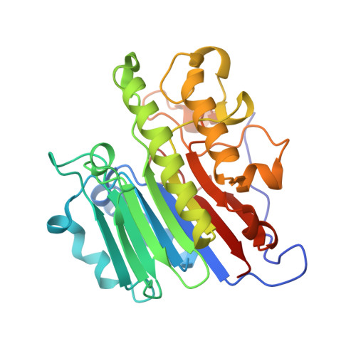
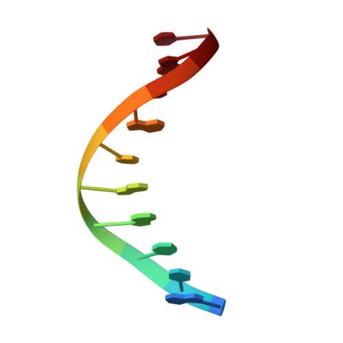
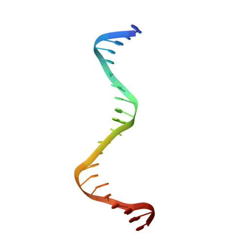
AP-endonuclease 1 sculpts DNA through an anchoring tyrosine residue on the DNA intercalating loop.

Hoitsma, N.M.Whitaker, A.M.Beckwitt, E.C.Jang, S.Agarwal, P.K.Van Houten, B.Freudenthal, B.D.

(2020) Nucleic Acids Res 48: 7345-7355

  • DOI: https://doi.org/10.1093/nar/gkaa496
  • Primary Citation of Related Structures:  
    6W4I, 6W4T

  • PubMed Abstract: 

    Base excision repair (BER) maintains genomic stability through the repair of DNA damage. Within BER, AP-endonuclease 1 (APE1) is a multifunctional enzyme that processes DNA intermediates through its backbone cleavage activity. To accomplish these repair activities, APE1 must recognize and accommodate several diverse DNA substrates. This is hypothesized to occur through a DNA sculpting mechanism where structural adjustments of the DNA substrate are imposed by the protein; however, how APE1 uniquely sculpts each substrate within a single rigid active site remains unclear. Here, we utilize structural and biochemical approaches to probe the DNA sculpting mechanism of APE1, specifically by characterizing a protein loop that intercalates the minor groove of the DNA (termed the intercalating loop). Pre-steady-state kinetics reveal a tyrosine residue within the intercalating loop (Y269) that is critical for AP-endonuclease activity. Using X-ray crystallography and molecular dynamics simulations, we determined the Y269 residue acts to anchor the intercalating loop on abasic DNA. Atomic force microscopy reveals the Y269 residue is required for proper DNA bending by APE1, providing evidence for the importance of this mechanism. We conclude that this previously unappreciated tyrosine residue is key to anchoring the intercalating loop and stabilizing the DNA in the APE1 active site.


  • Organizational Affiliation

    Department of Biochemistry and Molecular Biology, University of Kansas Medical Center, Kansas City, KS 66160, USA.


Macromolecules

Find similar proteins by:  (by identity cutoff)  |  3D Structure
Entity ID: 1
MoleculeChains Sequence LengthOrganismDetailsImage
DNA-(apurinic or apyrimidinic site) lyaseA,
E [auth B]
276Homo sapiensMutation(s): 2 
Gene Names: APEX1APEAPE1APEXAPXHAP1REF1
EC: 3.1 (PDB Primary Data), 4.2.99.18 (PDB Primary Data)
UniProt & NIH Common Fund Data Resources
Find proteins for P27695 (Homo sapiens)
Explore P27695 
Go to UniProtKB:  P27695
PHAROS:  P27695
GTEx:  ENSG00000100823 
Entity Groups  
Sequence Clusters30% Identity50% Identity70% Identity90% Identity95% Identity100% Identity
UniProt GroupP27695
Sequence Annotations
Expand
  • Reference Sequence

Find similar nucleic acids by:  Sequence   |   3D Structure  

Entity ID: 2
MoleculeChains LengthOrganismImage
DNA (5'-D(P*(3DR)P*CP*GP*AP*CP*GP*GP*AP*TP*CP*C)-3')B [auth D],
F [auth C]
11synthetic construct
Sequence Annotations
Expand
  • Reference Sequence

Find similar nucleic acids by:  Sequence   |   3D Structure  

Entity ID: 3
MoleculeChains LengthOrganismImage
DNA (5'-D(*GP*CP*TP*GP*AP*TP*GP*CP*GP*C)-3')C [auth P],
G [auth E]
10synthetic construct
Sequence Annotations
Expand
  • Reference Sequence

Find similar nucleic acids by:  Sequence   |   3D Structure  

Entity ID: 4
MoleculeChains LengthOrganismImage
DNA (5'-D(P*GP*GP*AP*TP*CP*CP*GP*TP*CP*GP*GP*GP*CP*GP*CP*AP*TP*CP*AP*GP*C)-3')D [auth V],
H [auth F]
21synthetic construct
Sequence Annotations
Expand
  • Reference Sequence
Experimental Data & Validation

Experimental Data

  • Method: X-RAY DIFFRACTION
  • Resolution: 2.20 Å
  • R-Value Free: 0.227 
  • R-Value Work: 0.185 
  • R-Value Observed: 0.187 
  • Space Group: P 21 21 21
Unit Cell:
Length ( Å )Angle ( ˚ )
a = 45.381α = 90
b = 150.233β = 90
c = 156.45γ = 90
Software Package:
Software NamePurpose
PHENIXrefinement
HKL-2000data collection
HKL-2000data scaling
PDB_EXTRACTdata extraction
HKL-2000data reduction
PHENIXphasing

Structure Validation

View Full Validation Report


Entry History & Funding Information

Deposition Data


Funding OrganizationLocationGrant Number
National Institutes of Health/National Institute of Environmental Health Sciences (NIH/NIEHS)United StatesR01 ES029203

Revision History  (Full details and data files)

  • Version 1.0: 2020-06-17
    Type: Initial release
  • Version 1.1: 2020-12-30
    Changes: Database references, Derived calculations
  • Version 1.2: 2023-10-18
    Changes: Data collection, Database references, Refinement description