6W2M

Abortive ternary complex crystal structure of DNA polymerase Beta with 8OG-dC base pair at the primer terminus and flipped out dA


Experimental Data Snapshot

  • Method: X-RAY DIFFRACTION
  • Resolution: 2.00 Å
  • R-Value Free: 0.238 
  • R-Value Work: 0.193 
  • R-Value Observed: 0.196 

wwPDB Validation   3D Report Full Report


Ligand Structure Quality Assessment 


This is version 1.1 of the entry. See complete history


Literature

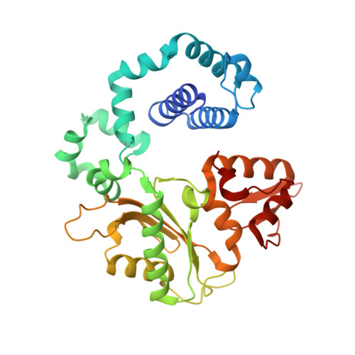
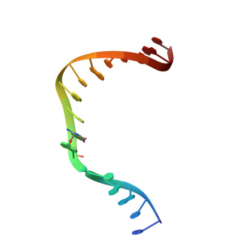
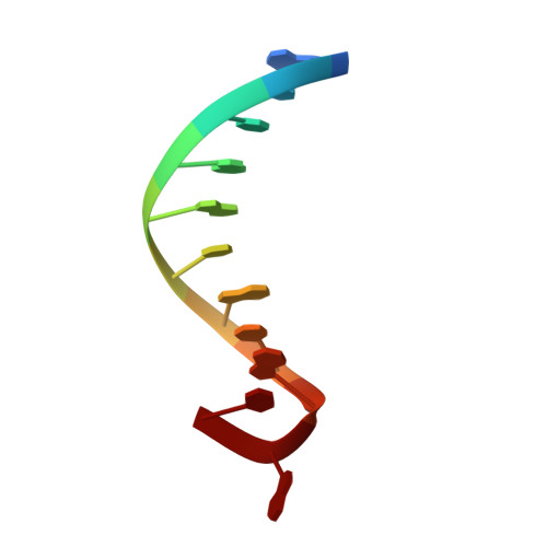
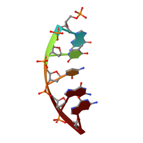
Structure of a DNA polymerase abortive complex with the 8OG:dA base pair at the primer terminus.

Batra, V.K.Wilson, S.H.

(2020) Commun Biol 3: 348-348

  • DOI: https://doi.org/10.1038/s42003-020-1080-4
  • Primary Citation of Related Structures:  
    6W2M

  • PubMed Abstract: 

    Adenine frequently pairs with the Hoogsteen edge of an oxidized guanine base (8OG) causing G to T transversions. The (syn)8OG:dA base pair is indistinguishable from the cognant base pair and can be extended by DNA polymerases with reduced efficiency. To examine the structural basis of this reduced efficiency, we sought to obtain the structure of the "product" complex of DNA polymerase (pol) β with the (syn)8OG:dA base pair at the primer terminus by soaking the binary complex crystals with a hydrolysable dCTP analogue complementary to the template base G. Crystallographic refinement of the structure revealed that the adenine of the (syn)8OG:dA base pair had been expelled from the primer terminus and a dCMP was inserted opposite 8OG in a reverse orientation; another uninserted molecule of the analogue was bound to the templating base G. This leads to an abortive complex that could form the basis of oxidatively-induced pol β stalling.


  • Organizational Affiliation

    Genome Integrity and Structural Biology Laboratory, National Institute of Environmental Health Sciences, National Institutes of Health, 111 T.W. Alexander Drive, Research Triangle Park, NC, 27709, USA.


Macromolecules

Find similar proteins by:  (by identity cutoff)  |  3D Structure
Entity ID: 1
MoleculeChains Sequence LengthOrganismDetailsImage
DNA polymerase beta335Homo sapiensMutation(s): 0 
Gene Names: POLB
EC: 2.7.7.7 (PDB Primary Data), 4.2.99 (PDB Primary Data)
UniProt & NIH Common Fund Data Resources
Find proteins for P06746 (Homo sapiens)
Explore P06746 
Go to UniProtKB:  P06746
PHAROS:  P06746
GTEx:  ENSG00000070501 
Entity Groups  
Sequence Clusters30% Identity50% Identity70% Identity90% Identity95% Identity100% Identity
UniProt GroupP06746
Sequence Annotations
Expand
  • Reference Sequence

Find similar nucleic acids by:  Sequence   |   3D Structure  

Entity ID: 2
MoleculeChains LengthOrganismImage
Template StrandB [auth T]16synthetic construct
Sequence Annotations
Expand
  • Reference Sequence

Find similar nucleic acids by:  Sequence   |   3D Structure  

Entity ID: 3
MoleculeChains LengthOrganismImage
Primer StrandC [auth P]11synthetic construct
Sequence Annotations
Expand
  • Reference Sequence

Find similar nucleic acids by:  Sequence   |   3D Structure  

Entity ID: 4
MoleculeChains LengthOrganismImage
Downstream Primer Strand5synthetic construct
Sequence Annotations
Expand
  • Reference Sequence
Small Molecules
Ligands 4 Unique
IDChains Name / Formula / InChI Key2D Diagram3D Interactions
SFV (Subject of Investigation/LOI)
Query on SFV

Download Ideal Coordinates CCD File 
K [auth A][[[(2~{R},3~{S},5~{R})-5-(4-azanyl-2-oxidanylidene-pyrimidin-1-yl)-3-oxidanyl-oxolan-2-yl]methoxy-oxidanyl-phosphoryl]oxy-oxidanyl-phosphoryl]methylphosphonic acid
C10 H18 N3 O12 P3
YHOBQJYELBYSJU-LKEWCRSYSA-N
CL
Query on CL

Download Ideal Coordinates CCD File 
I [auth A],
J [auth A]
CHLORIDE ION
Cl
VEXZGXHMUGYJMC-UHFFFAOYSA-M
MG
Query on MG

Download Ideal Coordinates CCD File 
E [auth A],
F [auth A]
MAGNESIUM ION
Mg
JLVVSXFLKOJNIY-UHFFFAOYSA-N
NA
Query on NA

Download Ideal Coordinates CCD File 
G [auth A],
H [auth A]
SODIUM ION
Na
FKNQFGJONOIPTF-UHFFFAOYSA-N
Experimental Data & Validation

Experimental Data

  • Method: X-RAY DIFFRACTION
  • Resolution: 2.00 Å
  • R-Value Free: 0.238 
  • R-Value Work: 0.193 
  • R-Value Observed: 0.196 
  • Space Group: P 1 21 1
Unit Cell:
Length ( Å )Angle ( ˚ )
a = 50.751α = 90
b = 80.462β = 107.75
c = 55.612γ = 90
Software Package:
Software NamePurpose
HKL-2000data reduction
PHENIXrefinement
PDB_EXTRACTdata extraction
HKL-2000data scaling
PHENIXphasing

Structure Validation

View Full Validation Report


Ligand Structure Quality Assessment 


Entry History 

Deposition Data

Revision History  (Full details and data files)

  • Version 1.0: 2020-07-22
    Type: Initial release
  • Version 1.1: 2023-10-18
    Changes: Data collection, Database references, Derived calculations, Refinement description