The first DEP domain of the RhoGEF P-Rex1 autoinhibits activity and contributes to membrane binding.
Ravala, S.K., Hopkins, J.B., Plescia, C.B., Allgood, S.R., Kane, M.A., Cash, J.N., Stahelin, R.V., Tesmer, J.J.G.(2020) J Biological Chem 295: 12635-12647
- PubMed: 32661198 Search on PubMedSearch on PubMed Central
- DOI: https://doi.org/10.1074/jbc.RA120.014534
- Primary Citation Related Structures:
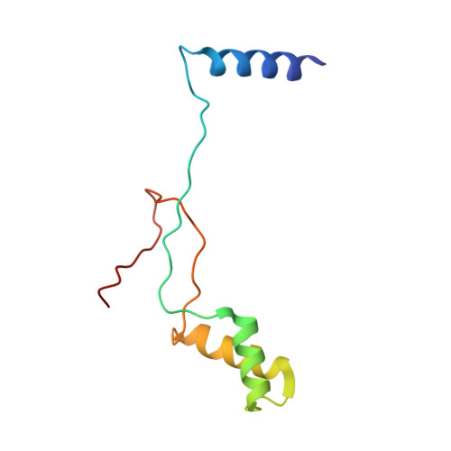
6VSK - PubMed Abstract:
Phosphatidylinositol (3,4,5)-trisphosphate (PIP 3 )-dependent Rac exchanger 1 (P-Rex1) catalyzes the exchange of GDP for GTP on Rac GTPases, thereby triggering changes in the actin cytoskeleton and in transcription. Its overexpression is highly correlated with the metastasis of certain cancers. P-Rex1 recruitment to the plasma membrane and its activity are regulated via interactions with heterotrimeric Gβγ subunits, PIP 3 , and protein kinase A (PKA). Deletion analysis has further shown that domains C-terminal to its catalytic Dbl homology (DH) domain confer autoinhibition. Among these, the first dishevelled, Egl-10, and pleckstrin domain (DEP1) remains to be structurally characterized. DEP1 also harbors the primary PKA phosphorylation site, suggesting that an improved understanding of this region could substantially increase our knowledge of P-Rex1 signaling and open the door to new selective chemotherapeutics. Here we show that the DEP1 domain alone can autoinhibit activity in context of the DH/PH-DEP1 fragment of P-Rex1 and interacts with the DH/PH domains in solution. The 3.1 Å crystal structure of DEP1 features a domain swap, similar to that observed previously in the Dvl2 DEP domain, involving an exposed basic loop that contains the PKA site. Using purified proteins, we show that although DEP1 phosphorylation has no effect on the activity or solution conformation of the DH/PH-DEP1 fragment, it inhibits binding of the DEP1 domain to liposomes containing phosphatidic acid. Thus, we propose that PKA phosphorylation of the DEP1 domain hampers P-Rex1 binding to negatively charged membranes in cells, freeing the DEP1 domain to associate with and inhibit the DH/PH module.
- Departments of Biological Sciences, Purdue University, West Lafayette, Indiana, USA.
Organizational Affiliation: