6VJW

Crystal structure of WT hMBD4 complexed with T:G mismatch DNA


Experimental Data Snapshot

  • Method: X-RAY DIFFRACTION
  • Resolution: 2.02 Å
  • R-Value Free: 0.263 
  • R-Value Work: 0.219 

wwPDB Validation   3D Report Full Report


This is version 1.1 of the entry. See complete history


Literature

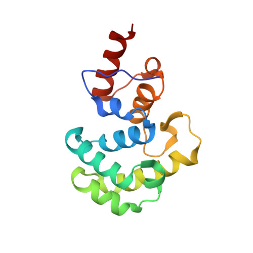
Catalytic mechanism of the mismatch-specific DNA glycosylase methyl-CpG-binding domain 4.

Ouzon-Shubeita, H.Jung, H.Lee, M.H.Koag, M.C.Lee, S.

(2020) Biochem J 477: 1601-1612

  • DOI: https://doi.org/10.1042/BCJ20200125
  • Primary Citation of Related Structures:  
    6VJW

  • PubMed Abstract: 

    Thymine:guanine base pairs are major promutagenic mismatches occurring in DNA metabolism. If left unrepaired, these mispairs can cause C to T transition mutations. In humans, T:G mismatches are repaired in part by mismatch-specific DNA glycosylases such as methyl-CpG-binding domain 4 (hMBD4) and thymine-DNA glycosylase. Unlike lesion-specific DNA glycosylases, T:G-mismatch-specific DNA glycosylases specifically recognize both bases of the mismatch and remove the thymine but only from mispairs with guanine. Despite the advances in biochemical and structural characterizations of hMBD4, the catalytic mechanism of hMBD4 remains elusive. Herein, we report two structures of hMBD4 processing T:G-mismatched DNA. A high-resolution crystal structure of Asp560Asn hMBD4-T:G complex suggests that hMBD4-mediated glycosidic bond cleavage occurs via a general base catalysis mechanism assisted by Asp560. A structure of wild-type hMBD4 encountering T:G-containing DNA shows the generation of an apurinic/apyrimidinic (AP) site bearing the C1'-(S)-OH. The inversion of the stereochemistry at the C1' of the AP-site indicates that a nucleophilic water molecule approaches from the back of the thymine substrate, suggesting a bimolecular displacement mechanism (SN2) for hMBD4-catalyzed thymine excision. The AP-site is stabilized by an extensive hydrogen bond network in the MBD4 catalytic site, highlighting the role of MBD4 in protecting the genotoxic AP-site.


  • Organizational Affiliation

    Division of Chemical Biology and Medicinal Chemistry, College of Pharmacy, The University of Texas at Austin, Austin, TX 78712, U.S.A.


Macromolecules

Find similar proteins by:  (by identity cutoff)  |  3D Structure
Entity ID: 1
MoleculeChains Sequence LengthOrganismDetailsImage
Methyl-CpG-binding domain protein 4138Homo sapiensMutation(s): 0 
Gene Names: MBD4MED1
EC: 3.2.2
UniProt & NIH Common Fund Data Resources
Find proteins for O95243 (Homo sapiens)
Explore O95243 
Go to UniProtKB:  O95243
PHAROS:  O95243
GTEx:  ENSG00000129071 
Entity Groups  
Sequence Clusters30% Identity50% Identity70% Identity90% Identity95% Identity100% Identity
UniProt GroupO95243
Sequence Annotations
Expand
  • Reference Sequence

Find similar nucleic acids by:  Sequence   |   3D Structure  

Entity ID: 2
MoleculeChains LengthOrganismImage
DNA (5'-D(*CP*CP*AP*GP*CP*GP*(ORP)P*GP*CP*AP*GP*C)-3')B [auth C]12Homo sapiens
Sequence Annotations
Expand
  • Reference Sequence

Find similar nucleic acids by:  Sequence   |   3D Structure  

Entity ID: 3
MoleculeChains LengthOrganismImage
DNA (5'-D(*GP*CP*TP*GP*CP*GP*CP*GP*CP*TP*GP*G)-3')C [auth D]12Homo sapiens
Sequence Annotations
Expand
  • Reference Sequence
Experimental Data & Validation

Experimental Data

  • Method: X-RAY DIFFRACTION
  • Resolution: 2.02 Å
  • R-Value Free: 0.263 
  • R-Value Work: 0.219 
  • Space Group: P 21 21 21
Unit Cell:
Length ( Å )Angle ( ˚ )
a = 40.35α = 90
b = 58.433β = 90
c = 101.142γ = 90
Software Package:
Software NamePurpose
PHENIXrefinement
HKL-2000data reduction
HKL-2000data scaling
MOLREPphasing

Structure Validation

View Full Validation Report


Entry History 

Deposition Data

Revision History  (Full details and data files)

  • Version 1.0: 2021-01-27
    Type: Initial release
  • Version 1.1: 2023-10-11
    Changes: Data collection, Database references, Refinement description