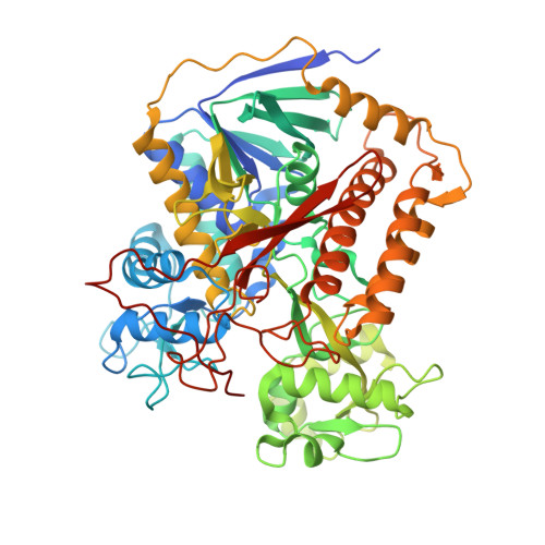
6VAX

Crystal structure of human SDHA-SDHAF2 assembly intermediate


Experimental Data Snapshot

  • Method: X-RAY DIFFRACTION
  • Resolution: 2.59 Å
  • R-Value Free: 0.249 
  • R-Value Work: 0.199 
  • R-Value Observed: 0.202 

Starting Models: experimental
View more details

wwPDB Validation   3D Report Full Report


Ligand Structure Quality Assessment 


This is version 1.5 of the entry. See complete history


Literature

The roles of SDHAF2 and dicarboxylate in covalent flavinylation of SDHA, the human complex II flavoprotein.

Sharma, P.Maklashina, E.Cecchini, G.Iverson, T.M.

(2020) Proc Natl Acad Sci U S A 117: 23548-23556

  • DOI: https://doi.org/10.1073/pnas.2007391117

  • PubMed Abstract: 

    Mitochondrial complex II, also known as succinate dehydrogenase (SDH), is an integral-membrane heterotetramer (SDHABCD) that links two essential energy-producing processes, the tricarboxylic acid (TCA) cycle and oxidative phosphorylation. A significant amount of information is available on the structure and function of mature complex II from a range of organisms. However, there is a gap in our understanding of how the enzyme assembles into a functional complex, and disease-associated complex II insufficiency may result from incorrect function of the mature enzyme or from assembly defects. Here, we investigate the assembly of human complex II by combining a biochemical reconstructionist approach with structural studies. We report an X-ray structure of human SDHA and its dedicated assembly factor SDHAF2. Importantly, we also identify a small molecule dicarboxylate that acts as an essential cofactor in this process and works in synergy with SDHAF2 to properly orient the flavin and capping domains of SDHA. This reorganizes the active site, which is located at the interface of these domains, and adjusts the pK a of SDHA R451 so that covalent attachment of the flavin adenine dinucleotide (FAD) cofactor is supported. We analyze the impact of disease-associated SDHA mutations on assembly and identify four distinct conformational forms of the complex II flavoprotein that we assign to roles in assembly and catalysis.


  • Organizational Affiliation

    Department of Pharmacology, Vanderbilt University, Nashville, TN 37232.


Macromolecules
Find similar proteins by:  (by identity cutoff)  |  3D Structure
Entity ID: 1
MoleculeChains Sequence LengthOrganismDetailsImage
Succinate dehydrogenase [ubiquinone] flavoprotein subunit, mitochondrial
A, C
632Homo sapiensMutation(s): 0 
Gene Names: SDHASDH2SDHF
EC: 1.3.5.1 (PDB Primary Data), 1.1.5 (UniProt)
UniProt & NIH Common Fund Data Resources
Find proteins for P31040 (Homo sapiens)
Explore P31040 
Go to UniProtKB:  P31040
PHAROS:  P31040
GTEx:  ENSG00000073578 
Entity Groups  
Sequence Clusters30% Identity50% Identity70% Identity90% Identity95% Identity100% Identity
UniProt GroupP31040
Sequence Annotations
Expand
  • Reference Sequence
Find similar proteins by:  (by identity cutoff)  |  3D Structure
Entity ID: 2
MoleculeChains Sequence LengthOrganismDetailsImage
Succinate dehydrogenase assembly factor 2, mitochondrial
B, D
166Homo sapiensMutation(s): 0 
Gene Names: SDHAF2C11orf79PGL2SDH5
UniProt & NIH Common Fund Data Resources
Find proteins for Q9NX18 (Homo sapiens)
Explore Q9NX18 
Go to UniProtKB:  Q9NX18
PHAROS:  Q9NX18
GTEx:  ENSG00000167985 
Entity Groups  
Sequence Clusters30% Identity50% Identity70% Identity90% Identity95% Identity100% Identity
UniProt GroupQ9NX18
Sequence Annotations
Expand
  • Reference Sequence
Small Molecules
Ligands 6 Unique
IDChains Name / Formula / InChI Key2D Diagram3D Interactions
FAD (Subject of Investigation/LOI)
Query on FAD

Download Ideal Coordinates CCD File 
E [auth A],
O [auth C]
FLAVIN-ADENINE DINUCLEOTIDE
C27 H33 N9 O15 P2
VWWQXMAJTJZDQX-UYBVJOGSSA-N
OAA (Subject of Investigation/LOI)
Query on OAA

Download Ideal Coordinates CCD File 
F [auth A],
P [auth C]
OXALOACETATE ION
C4 H3 O5
KHPXUQMNIQBQEV-UHFFFAOYSA-M
MLI
Query on MLI

Download Ideal Coordinates CCD File 
G [auth A]MALONATE ION
C3 H2 O4
OFOBLEOULBTSOW-UHFFFAOYSA-L
GOL
Query on GOL

Download Ideal Coordinates CCD File 
H [auth A],
N [auth C]
GLYCEROL
C3 H8 O3
PEDCQBHIVMGVHV-UHFFFAOYSA-N
EDO
Query on EDO

Download Ideal Coordinates CCD File 
J [auth A]
K [auth A]
L [auth A]
M [auth A]
R [auth C]
J [auth A],
K [auth A],
L [auth A],
M [auth A],
R [auth C],
S [auth C],
T [auth C],
U [auth C],
V [auth C]
1,2-ETHANEDIOL
C2 H6 O2
LYCAIKOWRPUZTN-UHFFFAOYSA-N
K
Query on K

Download Ideal Coordinates CCD File 
I [auth A],
Q [auth C]
POTASSIUM ION
K
NPYPAHLBTDXSSS-UHFFFAOYSA-N
Experimental Data & Validation

Experimental Data

Unit Cell:
Length ( Å )Angle ( ˚ )
a = 210.558α = 90
b = 68.782β = 93.16
c = 126.578γ = 90
Software Package:
Software NamePurpose
PHENIXrefinement
HKL-2000data reduction
HKL-2000data scaling
PHASERphasing

Structure Validation

View Full Validation Report


Ligand Structure Quality Assessment 


Entry History & Funding Information

Deposition Data


Funding OrganizationLocationGrant Number
National Institutes of Health/National Institute of General Medical Sciences (NIH/NIGMS)United StatesGM61606
American Heart AssociationUnited States19POST34450093

Revision History  (Full details and data files)

  • Version 1.0: 2020-08-26
    Type: Initial release
  • Version 1.1: 2020-09-16
    Changes: Database references
  • Version 1.2: 2020-10-07
    Changes: Database references
  • Version 1.3: 2023-03-08
    Changes: Database references, Structure summary
  • Version 1.4: 2023-10-25
    Changes: Data collection, Refinement description
  • Version 1.5: 2024-11-13
    Changes: Structure summary