6V7M

Crystal structure of a proteolytically cleaved, amino terminal domain of apolipoprotein E3


Experimental Data Snapshot

  • Method: X-RAY DIFFRACTION
  • Resolution: 2.00 Å
  • R-Value Free: 0.260 
  • R-Value Work: 0.203 
  • R-Value Observed: 0.205 

wwPDB Validation   3D Report Full Report

Currently 6V7M does not have a validation slider image.


This is version 1.1 of the entry. See complete history


Literature

Crystal structure of a proteolytically cleaved, amino terminal domain of apolipoprotein E3.

McPherson, A.

(2020) Biochem Biophys Res Commun 

  • DOI: https://doi.org/10.1016/j.bbrc.2020.01.117
  • Primary Citation of Related Structures:  
    6V7M

  • PubMed Abstract: 

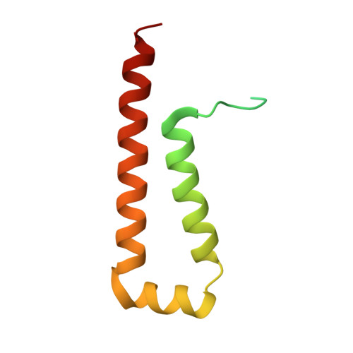
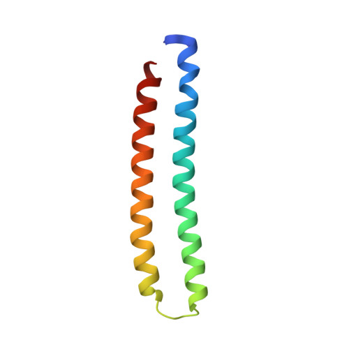
    A sample of Apolipoprotein E3 used in the original structure determination by X-ray crystallography (PDB code 1NFN) was crystallized under different conditions and its structure determined by molecular replacement at 298° K. The original model (1NFN) began at amino acid 23 and ended at amino acid 164, but the amino acid segment 81 through 91 (a loop between helices) was not visible in the electron density and presumed disordered. The model reported here is essentially identical to 1NFN, but now includes amino acids 18 through 22 at the amino terminus, 165 at the carboxy terminus and includes as well the segment 83 through 91. Leu 82 is not visible, but the separation between Gln 81 and Thr 83 is more than 10 Å, thereby indicating a proteolytic cleavage occurred between those two residues.


  • Organizational Affiliation

    Dept. Molecular Biology and Biochemistry, University of California, Irvine, 3205 McGaugh Hall Irvine, CA, 92697, USA. Electronic address: amcphers@uci.edu.


Macromolecules
Find similar proteins by:  (by identity cutoff)  |  3D Structure
Entity ID: 1
MoleculeChains Sequence LengthOrganismDetailsImage
Apolipoprotein E100Homo sapiensMutation(s): 0 
Gene Names: APOE
UniProt & NIH Common Fund Data Resources
Find proteins for P02649 (Homo sapiens)
Explore P02649 
Go to UniProtKB:  P02649
PHAROS:  P02649
GTEx:  ENSG00000130203 
Entity Groups  
Sequence Clusters30% Identity50% Identity70% Identity90% Identity95% Identity100% Identity
UniProt GroupP02649
Sequence Annotations
Expand
  • Reference Sequence
Find similar proteins by:  (by identity cutoff)  |  3D Structure
Entity ID: 2
MoleculeChains Sequence LengthOrganismDetailsImage
Apolipoprotein E83Homo sapiensMutation(s): 0 
Gene Names: APOE
UniProt & NIH Common Fund Data Resources
Find proteins for P02649 (Homo sapiens)
Explore P02649 
Go to UniProtKB:  P02649
PHAROS:  P02649
GTEx:  ENSG00000130203 
Entity Groups  
Sequence Clusters30% Identity50% Identity70% Identity90% Identity95% Identity100% Identity
UniProt GroupP02649
Sequence Annotations
Expand
  • Reference Sequence
Small Molecules
Ligands 1 Unique
IDChains Name / Formula / InChI Key2D Diagram3D Interactions
PO4
Query on PO4

Download Ideal Coordinates CCD File 
C [auth A]PHOSPHATE ION
O4 P
NBIIXXVUZAFLBC-UHFFFAOYSA-K
Experimental Data & Validation

Experimental Data

  • Method: X-RAY DIFFRACTION
  • Resolution: 2.00 Å
  • R-Value Free: 0.260 
  • R-Value Work: 0.203 
  • R-Value Observed: 0.205 
  • Space Group: P 21 21 21
Unit Cell:
Length ( Å )Angle ( ˚ )
a = 41.33α = 90
b = 54.45β = 90
c = 86.67γ = 90
Software Package:
Software NamePurpose
REFMACrefinement
PDB_EXTRACTdata extraction
SDMSdata reduction
Aimlessdata scaling
PHASERphasing

Structure Validation

View Full Validation Report

Currently 6V7M does not have a validation slider image.


Entry History 

Deposition Data

Revision History  (Full details and data files)

  • Version 1.0: 2020-03-11
    Type: Initial release
  • Version 1.1: 2023-10-11
    Changes: Data collection, Database references, Refinement description