Crystal structure of the nonclassical cadherin-17 N-terminus and implications for its adhesive binding mechanism.
Gray, M.E., Sotomayor, M.(2021) Acta Crystallogr F Struct Biol Commun 77: 85-94
- PubMed: 33682793 Search on PubMedSearch on PubMed Central
- DOI: https://doi.org/10.1107/S2053230X21002247
- Primary Citation Related Structures:
6ULM - PubMed Abstract:
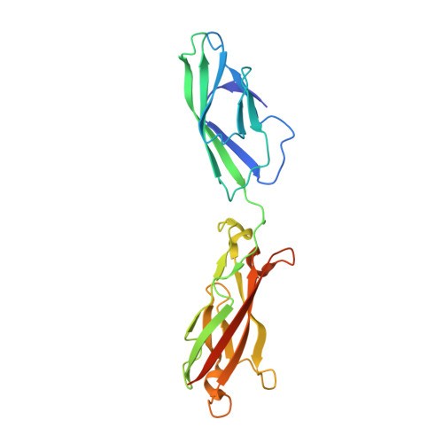
The cadherin superfamily of calcium-dependent cell-adhesion proteins has over 100 members in the human genome. All members of the superfamily feature at least a pair of extracellular cadherin (EC) repeats with calcium-binding sites in the EC linker region. The EC repeats across family members form distinct complexes that mediate cellular adhesion. For instance, classical cadherins (five EC repeats) strand-swap their N-termini and exchange tryptophan residues in EC1, while the clustered protocadherins (six EC repeats) use an extended antiparallel `forearm handshake' involving repeats EC1-EC4. The 7D-cadherins, cadherin-16 (CDH16) and cadherin-17 (CDH17), are the most similar to classical cadherins and have seven EC repeats, two of which are likely to have arisen from gene duplication of EC1-2 from a classical ancestor. However, CDH16 and CDH17 lack the EC1 tryptophan residue used by classical cadherins to mediate adhesion. The structure of human CDH17 EC1-2 presented here reveals features that are not seen in classical cadherins and that are incompatible with the EC1 strand-swap mechanism for adhesion. Analyses of crystal contacts, predicted glycosylation and disease-related mutations are presented along with sequence alignments suggesting that the novel features in the CDH17 EC1-2 structure are well conserved. These results hint at distinct adhesive properties for 7D-cadherins.
- Department of Chemistry and Biochemistry, The Ohio State University, 484 West 12th Avenue, Columbus, OH 43210, USA.
Organizational Affiliation: