6UIW

Cryo-EM structure of human CALHM2 in a ruthenium red-bound inhibited state


Experimental Data Snapshot

  • Method: ELECTRON MICROSCOPY
  • Resolution: 2.70 Å
  • Aggregation State: PARTICLE 
  • Reconstruction Method: SINGLE PARTICLE 

wwPDB Validation   3D Report Full Report


This is version 1.3 of the entry. See complete history


Literature

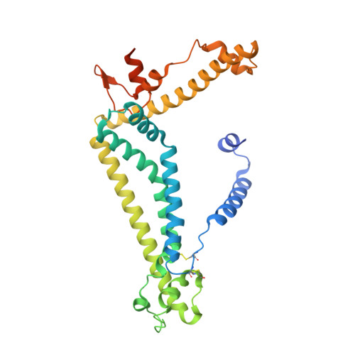
The structures and gating mechanism of human calcium homeostasis modulator 2.

Choi, W.Clemente, N.Sun, W.Du, J.Lu, W.

(2019) Nature 576: 163-167

  • DOI: https://doi.org/10.1038/s41586-019-1781-3
  • Primary Citation of Related Structures:  
    6UIV, 6UIW, 6UIX

  • PubMed Abstract: 

    Calcium homeostasis modulators (CALHMs) are voltage-gated, Ca 2+ -inhibited nonselective ion channels that act as major ATP release channels, and have important roles in gustatory signalling and neuronal toxicity 1-3 . Dysfunction of CALHMs has previously been linked to neurological disorders 1 . Here we present cryo-electron microscopy structures of the human CALHM2 channel in the Ca 2+ -free active or open state and in the ruthenium red (RUR)-bound inhibited state, at resolutions up to 2.7 Å. Our work shows that purified CALHM2 channels form both gap junctions and undecameric hemichannels. The protomer shows a mirrored arrangement of the transmembrane domains (helices S1-S4) relative to other channels with a similar topology, such as connexins, innexins and volume-regulated anion channels 4-8 . Upon binding to RUR, we observed a contracted pore with notable conformational changes of the pore-lining helix S1, which swings nearly 60° towards the pore axis from a vertical to a lifted position. We propose a two-section gating mechanism in which the S1 helix coarsely adjusts, and the N-terminal helix fine-tunes, the pore size. We identified a RUR-binding site near helix S1 that may stabilize this helix in the lifted conformation, giving rise to channel inhibition. Our work elaborates on the principles of CALHM2 channel architecture and symmetry, and the mechanism that underlies channel inhibition.


  • Organizational Affiliation

    Van Andel Institute, Grand Rapids, MI, USA.


Macromolecules
Find similar proteins by:  (by identity cutoff)  |  3D Structure
Entity ID: 1
MoleculeChains Sequence LengthOrganismDetailsImage
Calcium homeostasis modulator protein 2
A, B, C, D, E
A, B, C, D, E, F, G, H, I, J, K
331Homo sapiensMutation(s): 0 
Gene Names: CALHM2FAM26B
Membrane Entity: Yes 
UniProt & NIH Common Fund Data Resources
Find proteins for Q9HA72 (Homo sapiens)
Explore Q9HA72 
Go to UniProtKB:  Q9HA72
PHAROS:  Q9HA72
GTEx:  ENSG00000138172 
Entity Groups  
Sequence Clusters30% Identity50% Identity70% Identity90% Identity95% Identity100% Identity
UniProt GroupQ9HA72
Sequence Annotations
Expand
  • Reference Sequence
Small Molecules
Ligands 1 Unique
IDChains Name / Formula / InChI Key2D Diagram3D Interactions
R2R (Subject of Investigation/LOI)
Query on R2R

Download Ideal Coordinates CCD File 
L [auth A]
M [auth B]
N [auth C]
O [auth D]
P [auth E]
L [auth A],
M [auth B],
N [auth C],
O [auth D],
P [auth E],
Q [auth F],
R [auth G],
S [auth H],
T [auth I],
U [auth J],
V [auth K]
ruthenium(6+) azanide pentaamino(oxido)ruthenium (1/4/2)
H28 N14 O2 Ru3
FOOPNNMQSUSGAT-UHFFFAOYSA-N
Experimental Data & Validation

Experimental Data

  • Method: ELECTRON MICROSCOPY
  • Resolution: 2.70 Å
  • Aggregation State: PARTICLE 
  • Reconstruction Method: SINGLE PARTICLE 
EM Software:
TaskSoftware PackageVersion
RECONSTRUCTIONRELION3.0
MODEL REFINEMENTPHENIX1.16

Structure Validation

View Full Validation Report


Entry History 

Deposition Data

Revision History  (Full details and data files)

  • Version 1.0: 2019-11-27
    Type: Initial release
  • Version 1.1: 2019-12-04
    Changes: Structure summary
  • Version 1.2: 2019-12-11
    Changes: Database references
  • Version 1.3: 2019-12-18
    Changes: Database references