Expression of quasi-equivalence and capsid dimorphism in the Hepadnaviridae.
Wu, W., Watts, N.R., Cheng, N., Huang, R., Steven, A.C., Wingfield, P.T.(2020) PLoS Comput Biol 16: e1007782-e1007782
- PubMed: 32310951
- DOI: https://doi.org/10.1371/journal.pcbi.1007782
- Primary Citation Related Structures:
6UI6, 6UI7 - PubMed Abstract:
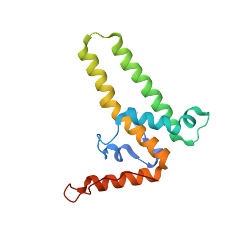
Hepatitis B virus (HBV) is a leading cause of liver disease. The capsid is an essential component of the virion and it is therefore of interest how it assembles and disassembles. The capsid protein is unusual both for its rare fold and that it polymerizes according to two different icosahedral symmetries, causing the polypeptide chain to exist in seven quasi-equivalent environments: A, B, and C in AB and CC dimers in T = 3 capsids, and A, B, C, and D in AB and CD dimers in T = 4 capsids. We have compared the two capsids by cryo-EM at 3.5 Å resolution. To ensure a valid comparison, the two capsids were prepared and imaged under identical conditions. We find that the chains have different conformations and potential energies, with the T = 3 C chain having the lowest. Three of the four quasi-equivalent dimers are asymmetric with respect to conformation and potential energy; however, the T = 3 CC dimer is symmetrical and has the lowest potential energy although its intra-dimer interface has the least free energy of formation. Of all the inter-dimer interfaces, the CB interface has the least area and free energy, in both capsids. From the calculated energies of higher-order groupings of dimers discernible in the lattices we predict early assembly intermediates, and indeed we observe such structures by negative stain EM of in vitro assembly reactions. By sequence analysis and computational alanine scanning we identify key residues and motifs involved in capsid assembly. Our results explain several previously reported observations on capsid assembly, disassembly, and dimorphism.
- Laboratory of Structural Biology Research, National Institute of Arthritis and Musculoskeletal and Skin Diseases, National Institutes of Health, Bethesda, Maryland, United States of America.
Organizational Affiliation: