6TZU

Dihydrodipicolinate synthase (DHDPS) from C.jejuni, N84A mutant with pyruvate bound in the active site


Experimental Data Snapshot

  • Method: X-RAY DIFFRACTION
  • Resolution: 1.80 Å
  • R-Value Free: 0.201 
  • R-Value Work: 0.172 
  • R-Value Observed: 0.174 

wwPDB Validation   3D Report Full Report


This is version 1.3 of the entry. See complete history


Literature

Asparagine-84, a regulatory allosteric site residue, helps maintain the quaternary structure of Campylobacter jejuni dihydrodipicolinate synthase.

Majdi Yazdi, M.Saran, S.Mrozowich, T.Lehnert, C.Patel, T.R.Sanders, D.A.R.Palmer, D.R.J.

(2020) J Struct Biol 209: 107409-107409

  • DOI: https://doi.org/10.1016/j.jsb.2019.107409
  • Primary Citation of Related Structures:  
    6TZU, 6U01

  • PubMed Abstract: 

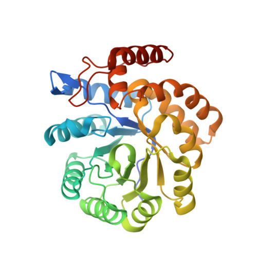
    Dihydrodipicolinate synthase (DHDPS) from Campylobacter jejuni is a natively homotetrameric enzyme that catalyzes the first unique reaction of (S)-lysine biosynthesis and is feedback-regulated by lysine through binding to an allosteric site. High-resolution structures of the DHDPS-lysine complex have revealed significant insights into the binding events. One key asparagine residue, N84, makes hydrogen bonds with both the carboxyl and the α-amino group of the bound lysine. We generated two mutants, N84A and N84D, to study the effects of these changes on the allosteric site properties. However, under normal assay conditions, N84A displayed notably lower catalytic activity, and N84D showed no activity. Here we show that these mutations disrupt the quaternary structure of DHDPS in a concentration-dependent fashion, as demonstrated by size-exclusion chromatography, multi-angle light scattering, dynamic light scattering, small-angle X-ray scattering (SAXS) and high-resolution protein crystallography.


  • Organizational Affiliation

    Department of Chemistry, University of Saskatchewan, 110 Science Place, Saskatoon, SK S7N 5C9, Canada.


Macromolecules
Find similar proteins by:  (by identity cutoff)  |  3D Structure
Entity ID: 1
MoleculeChains Sequence LengthOrganismDetailsImage
4-hydroxy-tetrahydrodipicolinate synthase
A, B, C, D, E
310Campylobacter jejuni subsp. jejuni NCTC 11168 = ATCC 700819Mutation(s): 1 
Gene Names: dapACj0806
EC: 4.3.3.7
UniProt
Find proteins for Q9PPB4 (Campylobacter jejuni subsp. jejuni serotype O:2 (strain ATCC 700819 / NCTC 11168))
Explore Q9PPB4 
Go to UniProtKB:  Q9PPB4
Entity Groups  
Sequence Clusters30% Identity50% Identity70% Identity90% Identity95% Identity100% Identity
UniProt GroupQ9PPB4
Sequence Annotations
Expand
  • Reference Sequence
Find similar proteins by:  (by identity cutoff)  |  3D Structure
Entity ID: 2
MoleculeChains Sequence LengthOrganismDetailsImage
4-hydroxy-tetrahydrodipicolinate synthase310Campylobacter jejuni subsp. jejuni NCTC 11168 = ATCC 700819Mutation(s): 1 
Gene Names: dapACj0806
EC: 4.3.3.7
UniProt
Find proteins for Q9PPB4 (Campylobacter jejuni subsp. jejuni serotype O:2 (strain ATCC 700819 / NCTC 11168))
Explore Q9PPB4 
Go to UniProtKB:  Q9PPB4
Entity Groups  
Sequence Clusters30% Identity50% Identity70% Identity90% Identity95% Identity100% Identity
UniProt GroupQ9PPB4
Sequence Annotations
Expand
  • Reference Sequence
Small Molecules
Ligands 6 Unique
IDChains Name / Formula / InChI Key2D Diagram3D Interactions
PGE
Query on PGE

Download Ideal Coordinates CCD File 
IB [auth F],
N [auth A],
NA [auth D],
Y [auth B]
TRIETHYLENE GLYCOL
C6 H14 O4
ZIBGPFATKBEMQZ-UHFFFAOYSA-N
PEG
Query on PEG

Download Ideal Coordinates CCD File 
EA [auth C],
FA [auth C],
X [auth B],
YA [auth E]
DI(HYDROXYETHYL)ETHER
C4 H10 O3
MTHSVFCYNBDYFN-UHFFFAOYSA-N
GOL
Query on GOL

Download Ideal Coordinates CCD File 
OA [auth D],
ZA [auth E]
GLYCEROL
C3 H8 O3
PEDCQBHIVMGVHV-UHFFFAOYSA-N
EDO
Query on EDO

Download Ideal Coordinates CCD File 
BB [auth F]
CB [auth F]
DB [auth F]
GA [auth D]
HA [auth D]
BB [auth F],
CB [auth F],
DB [auth F],
GA [auth D],
HA [auth D],
I [auth A],
IA [auth D],
J [auth A],
JA [auth D],
K [auth A],
KA [auth D],
L [auth A],
LA [auth D],
MA [auth D],
P [auth B],
Q [auth B],
QA [auth E],
R [auth B],
RA [auth E],
S [auth B],
SA [auth E],
T [auth B],
TA [auth E],
U [auth B]
1,2-ETHANEDIOL
C2 H6 O2
LYCAIKOWRPUZTN-UHFFFAOYSA-N
ACT
Query on ACT

Download Ideal Coordinates CCD File 
AA [auth C]
BA [auth C]
CA [auth C]
DA [auth C]
EB [auth F]
AA [auth C],
BA [auth C],
CA [auth C],
DA [auth C],
EB [auth F],
FB [auth F],
GB [auth F],
HB [auth F],
M [auth A],
UA [auth E],
V [auth B],
VA [auth E],
W [auth B],
WA [auth E],
XA [auth E],
Z [auth C]
ACETATE ION
C2 H3 O2
QTBSBXVTEAMEQO-UHFFFAOYSA-M
MG
Query on MG

Download Ideal Coordinates CCD File 
AB [auth F],
G [auth A],
H [auth A],
O [auth B],
PA [auth E]
MAGNESIUM ION
Mg
JLVVSXFLKOJNIY-UHFFFAOYSA-N
Modified Residues  1 Unique
IDChains TypeFormula2D DiagramParent
KPI
Query on KPI
F
L-PEPTIDE LINKINGC9 H16 N2 O4LYS
Experimental Data & Validation

Experimental Data

  • Method: X-RAY DIFFRACTION
  • Resolution: 1.80 Å
  • R-Value Free: 0.201 
  • R-Value Work: 0.172 
  • R-Value Observed: 0.174 
  • Space Group: C 2 2 21
Unit Cell:
Length ( Å )Angle ( ˚ )
a = 84.93α = 90
b = 225.81β = 90
c = 200.74γ = 90
Software Package:
Software NamePurpose
Cootmodel building
PHENIXrefinement
XSCALEdata scaling
HKL-3000data collection
PHENIXphasing

Structure Validation

View Full Validation Report


Entry History & Funding Information

Deposition Data


Funding OrganizationLocationGrant Number
Natural Sciences and Engineering Research Council (NSERC, Canada)Canada--

Revision History  (Full details and data files)

  • Version 1.0: 2019-12-04
    Type: Initial release
  • Version 1.1: 2020-01-22
    Changes: Database references
  • Version 1.2: 2023-10-11
    Changes: Data collection, Database references, Derived calculations, Refinement description
  • Version 1.3: 2023-11-29
    Changes: Data collection