6TUK | pdb_00006tuk

Crystal structure of Fdr9


Experimental Data Snapshot

  • Method: X-RAY DIFFRACTION
  • Resolution: 1.90 Å
  • R-Value Free: 
    0.199 (Depositor), 0.199 (DCC) 
  • R-Value Work: 
    0.184 (Depositor), 0.185 (DCC) 
  • R-Value Observed: 
    0.185 (Depositor) 

Starting Model: experimental
View more details

wwPDB Validation 3D Report Full Report

Validation slider image for 6TUK

Ligand Structure Quality Assessment 


This is version 1.1 of the entry. See complete history

Literature

Expression, purification and crystal structure determination of a ferredoxin reductase from the actinobacterium Thermobifida fusca.

Rodriguez Buitrago, J.A.Klunemann, T.Blankenfeldt, W.Schallmey, A.

(2020) Acta Crystallogr F Struct Biol Commun 76: 334-340

  • DOI: https://doi.org/10.1107/S2053230X2000922X
  • Primary Citation Related Structures: 
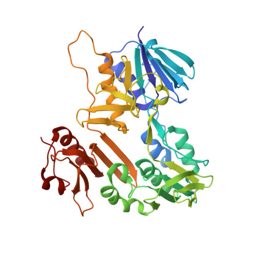
    6TUK

  • PubMed Abstract: 

    The ferredoxin reductase FdR9 from Thermobifida fusca, a member of the oxygenase-coupled NADH-dependent ferredoxin reductase (FNR) family, catalyses electron transfer from NADH to its physiological electron acceptor ferredoxin. It forms part of a putative three-component cytochrome P450 monooxygenase system in T. fusca comprising CYP222A1 and the [3Fe-4S]-cluster ferredoxin Fdx8 as well as FdR9. Here, FdR9 was overexpressed and purified and its crystal structure was determined at 1.9 Å resolution. The overall structure of FdR9 is similar to those of other members of the FNR family and is composed of an FAD-binding domain, an NAD-binding domain and a C-terminal domain. Activity measurements with FdR9 confirmed a strong preference for NADH as the cofactor. Comparison of the FAD- and NAD-binding domains of FdR9 with those of other ferredoxin reductases revealed the presence of conserved sequence motifs in the FAD-binding domain as well as several highly conserved residues involved in FAD and NAD cofactor binding. Moreover, the NAD-binding site of FdR9 contains a modified Rossmann-fold motif, GxSxxS, instead of the classical GxGxxG motif.


  • Organizational Affiliation
    • Institute for Biochemistry, Biotechnology and Bioinformatics, Technical University Braunschweig, Spielmannstrasse 7, 38106 Braunschweig, Germany.

Macromolecule Content 

  • Total Structure Weight: 43.59 kDa 
  • Atom Count: 3,273 
  • Modeled Residue Count: 393 
  • Deposited Residue Count: 393 
  • Unique protein chains: 1

Macromolecules

Find similar proteins by:|  3D Structure
Entity ID: 1
MoleculeChains  Sequence LengthOrganismDetailsImage
Putative oxidoreductaseA [auth B]393Thermobifida fusca YXMutation(s): 0 
Gene Names: Tfu_1273
UniProt
Find proteins for Q47QF8 (Thermobifida fusca (strain YX))
Explore Q47QF8 
Go to UniProtKB:  Q47QF8
Entity Groups
Sequence Clusters30% Identity50% Identity70% Identity90% Identity95% Identity100% Identity
UniProt GroupQ47QF8
Sequence Annotations
Expand
Reference Sequence

Small Molecules

Ligands 3 Unique
IDChains Name / Formula / InChI Key2D Diagram3D Interactions
FAD

Query on FAD


Download:Ideal Coordinates CCD File
B
FLAVIN-ADENINE DINUCLEOTIDE
C27 H33 N9 O15 P2
VWWQXMAJTJZDQX-UYBVJOGSSA-N
SO4

Query on SO4


Download:Ideal Coordinates CCD File
F [auth B],
G [auth B],
H [auth B],
I [auth B]
SULFATE ION
O4 S
QAOWNCQODCNURD-UHFFFAOYSA-L
BU3

Query on BU3


Download:Ideal Coordinates CCD File
C [auth B],
D [auth B],
E [auth B]
(R,R)-2,3-BUTANEDIOL
C4 H10 O2
OWBTYPJTUOEWEK-QWWZWVQMSA-N

Experimental Data & Validation

Experimental Data

  • Method: X-RAY DIFFRACTION
  • Resolution: 1.90 Å
  • R-Value Free:  0.199 (Depositor), 0.199 (DCC) 
  • R-Value Work:  0.184 (Depositor), 0.185 (DCC) 
  • R-Value Observed: 0.185 (Depositor) 
Space Group: P 41 3 2
Unit Cell:
Length ( Å )Angle ( ˚ )
a = 142.791α = 90
b = 142.791β = 90
c = 142.791γ = 90
Software Package:
Software NamePurpose
Aimlessdata scaling
PHENIXrefinement
PDB_EXTRACTdata extraction
DIALSdata reduction
MrBUMPphasing

Structure Validation

View Full Validation Report


Ligand Structure Quality Assessment 


Entry History 

& Funding Information

Deposition Data


Funding OrganizationLocationGrant Number
German Research Foundation (DFG)GermanyGRK2223

Revision History  (Full details and data files)

  • Version 1.0: 2020-11-18
    Type: Initial release
  • Version 1.1: 2024-01-24
    Changes: Data collection, Database references, Refinement description