6TNE | pdb_00006tne

Crystal structure of receiver domain from Hybrid Histidine Kinase CckA


Experimental Data Snapshot

  • Method: X-RAY DIFFRACTION
  • Resolution: 1.25 Å
  • R-Value Free: 
    0.188 (Depositor), 0.188 (DCC) 
  • R-Value Work: 
    0.165 (Depositor), 0.165 (DCC) 
  • R-Value Observed: 
    0.166 (Depositor) 

Starting Model: other
View more details

wwPDB Validation 3D Report Full Report

Validation slider image for 6TNE

This is version 2.2 of the entry. See complete history

Literature

Structural features discriminating hybrid histidine kinase Rec domains from response regulator homologs.

Bruderlin, M.Bohm, R.Fadel, F.Hiller, S.Schirmer, T.Dubey, B.N.

(2023) Nat Commun 14: 1002-1002

  • DOI: https://doi.org/10.1038/s41467-023-36597-8
  • Primary Citation Related Structures: 
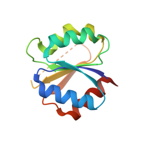
    6TNE

  • PubMed Abstract: 

    In two-component systems, the information gathered by histidine kinases (HKs) are relayed to cognate response regulators (RRs). Thereby, the phosphoryl group of the auto-phosphorylated HK is transferred to the receiver (Rec) domain of the RR to allosterically activate its effector domain. In contrast, multi-step phosphorelays comprise at least one additional Rec (Rec inter ) domain that is typically part of the HK and acts as an intermediary for phosphoryl-shuttling. While RR Rec domains have been studied extensively, little is known about discriminating features of Rec inter domains. Here we study the Rec inter domain of the hybrid HK CckA by X-ray crystallography and NMR spectroscopy. Strikingly, all active site residues of the canonical Rec-fold are pre-arranged for phosphoryl-binding and BeF 3 - binding does not alter secondary or quaternary structure, indicating the absence of allosteric changes, the hallmark of RRs. Based on sequence-covariation and modeling, we analyze the intra-molecular DHp/Rec association in hybrid HKs.


  • Organizational Affiliation
    • Structural Biology, Biozentrum, University of Basel, Spitalstr. 41, 4056, Basel, Switzerland.

Macromolecule Content 

  • Total Structure Weight: 12.96 kDa 
  • Atom Count: 943 
  • Modeled Residue Count: 108 
  • Deposited Residue Count: 120 
  • Unique protein chains: 1

Macromolecules

Find similar proteins by:|  3D Structure
Entity ID: 1
MoleculeChains  Sequence LengthOrganismDetailsImage
Histidine kinase120Caulobacter vibrioidesMutation(s): 0 
Gene Names: cckA
EC: 2.7.13.3
UniProt
Find proteins for Q9X688 (Caulobacter vibrioides)
Explore Q9X688 
Go to UniProtKB:  Q9X688
Entity Groups
Sequence Clusters30% Identity50% Identity70% Identity90% Identity95% Identity100% Identity
UniProt GroupQ9X688
Sequence Annotations
Expand
Reference Sequence

Experimental Data & Validation

Experimental Data

  • Method: X-RAY DIFFRACTION
  • Resolution: 1.25 Å
  • R-Value Free:  0.188 (Depositor), 0.188 (DCC) 
  • R-Value Work:  0.165 (Depositor), 0.165 (DCC) 
  • R-Value Observed: 0.166 (Depositor) 
Space Group: I 1 2 1
Unit Cell:
Length ( Å )Angle ( ˚ )
a = 37.43α = 90
b = 42.9β = 94.36
c = 65.037γ = 90
Software Package:
Software NamePurpose
PHENIXrefinement
MOSFLMdata reduction
Aimlessdata scaling
MOLREPphasing
PDB_EXTRACTdata extraction

Structure Validation

View Full Validation Report


Entry History 

& Funding Information

Deposition Data


Funding OrganizationLocationGrant Number
Swiss National Science FoundationSwitzerland31003A-166652

Revision History  (Full details and data files)

  • Version 1.0: 2020-12-16
    Type: Initial release
  • Version 2.0: 2022-10-05
    Type: Coordinate replacement
    Reason: Model completeness
    Changes: Advisory, Atomic model, Data collection, Database references, Derived calculations, Non-polymer description, Other, Polymer sequence, Refinement description, Source and taxonomy, Structure summary
  • Version 2.1: 2024-05-01
    Changes: Data collection, Refinement description
  • Version 2.2: 2025-09-10
    Changes: Database references, Structure summary