Structure and Multitasking of the c-di-GMP-Sensing Cellulose Secretion Regulator BcsE.
Zouhir, S., Abidi, W., Caleechurn, M., Krasteva, P.V.(2020) mBio 11
- PubMed: 32788377 Search on PubMedSearch on PubMed Central
- DOI: https://doi.org/10.1128/mBio.01303-20
- Primary Citation Related Structures:
6TJ0 - PubMed Abstract:
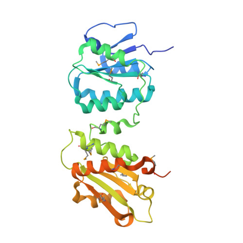
Most bacteria respond to surfaces by biogenesis of intracellular c-di-GMP, which inhibits motility and induces secretion of biofilm-promoting adherence factors. Bacterial cellulose is a widespread biofilm component whose secretion in Gram-negative species requires an inner membrane, c-di-GMP-dependent synthase tandem (BcsAB), an outer membrane porin (BcsC), and various accessory subunits that regulate synthase assembly and function as well as the exopolysaccharide's chemical composition and mechanical properties. We recently showed that in Escherichia coli , most Bcs proteins form a megadalton-sized secretory nanomachine, but the role and structure of individual regulatory components remained enigmatic. Here, we demonstrate that essential-for-secretion BcsR and BcsQ regulate each other's folding and stability and are recruited to the inner membrane via c-di-GMP-sensing BcsE and its intraoperon partner BcsF. Crystallographic and solution-based data show that BcsE's predicted GIL domain is a degenerate receiver-GGDEF domain tandem (BcsE REC * -GGDEF *), where the divergent diguanylate cyclase module binds both dimeric c-di-GMP and BcsQ through mutually independent interfaces. In addition, we reveal that a third N-terminal domain (BcsE NTD ) determines the protein's homooligomerization and targeting of BcsERQ to the membrane as well as previously unreported interactions with transcription antitermination complex components. Together, the data suggest that BcsE acts on multiple levels to fine-tune bacterial cellulose secretion, from the early stages of secretion system assembly to the maintenance of a membrane-proximal pool of dimeric c-di-GMP for processive synthase activation. IMPORTANCE Bacterial cellulose is a widespread biofilm component that can modulate microbial fitness and virulence both in the environment and infected hosts. Whereas its secretion generally involves an inner membrane c-di-GMP-dependent synthase tandem (BcsAB) across the bacterial domain of life, enterobacteria feature sophisticated Escherichia coli -like Bcs secretion systems, where multiple additional subunits are either required for secretion or contribute to the maximal production of the polysaccharide in vivo. Here, we demonstrate that essential-for-secretion BcsR and BcsQ regulate each other's folding and stability and are recruited to the inner membrane via c-di-GMP-sensing BcsE and its intraoperon partner, BcsF. Crystallographic and functional data reveal that BcsE features unexpected domain architecture and likely acts on multiple levels to fine-tune bacterial cellulose production, from the early stages of secretion system assembly to the maintenence of a membrane-proximal pool of dimeric c-di-GMP for processive synthase activation.
- Structural Biology of Biofilms Group, Institute for Integrative Biology of the Cell (I2BC), CEA, CNRS, Paris-Sud University, Gif-sur-Yvette, France.
Organizational Affiliation: