6SZ6

Chaetomium thermophilum beta-glucosidase


Experimental Data Snapshot

  • Method: X-RAY DIFFRACTION
  • Resolution: 2.99 Å
  • R-Value Free: 0.249 
  • R-Value Work: 0.201 
  • R-Value Observed: 0.203 

wwPDB Validation   3D Report Full Report


Ligand Structure Quality Assessment 


This is version 2.2 of the entry. See complete history


Literature

Crystal Structure of a GH3 beta-Glucosidase from the Thermophilic Fungus Chaetomium thermophilum .

Mohsin, I.Poudel, N.Li, D.C.Papageorgiou, A.C.

(2019) Int J Mol Sci 20

  • DOI: https://doi.org/10.3390/ijms20235962
  • Primary Citation of Related Structures:  
    6SZ6

  • PubMed Abstract: 

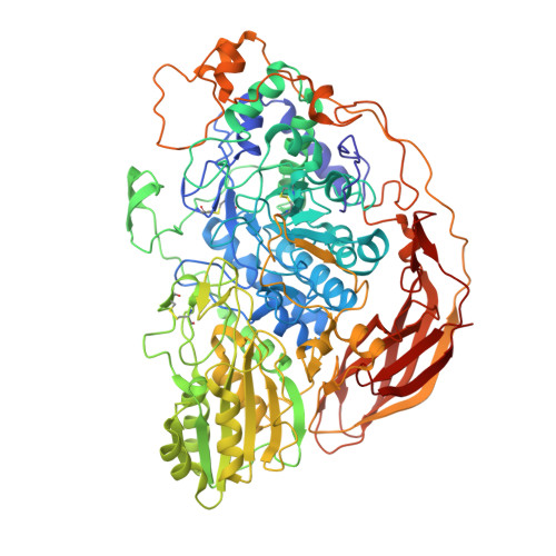
    Beta-glucosidases (β-glucosidases) have attracted considerable attention in recent years for use in various biotechnological applications. They are also essential enzymes for lignocellulose degradation in biofuel production. However, cost-effective biomass conversion requires the use of highly efficient enzymes. Thus, the search for new enzymes as better alternatives of the currently available enzyme preparations is highly important. Thermophilic fungi are nowadays considered as a promising source of enzymes with improved stability. Here, the crystal structure of a family GH3 β-glucosidase from the thermophilic fungus Chaetomium thermophilum ( Ct BGL) was determined at a resolution of 2.99 Å. The structure showed the three-domain architecture found in other β-glucosidases with variations in loops and linker regions. The active site catalytic residues in Ct BGL were identified as Asp287 (nucleophile) and Glu517 (acid/base). Structural comparison of Ct BGL with Protein Data Bank (PDB)-deposited structures revealed variations among glycosylated Asn residues. The enzyme displayed moderate glycosylation compared to other GH3 family β-glucosidases with similar structure. A new glycosylation site at position Asn504 was identified in Ct BGL. Moreover, comparison with respect to several thermostability parameters suggested that glycosylation and charged residues involved in electrostatic interactions may contribute to the stability of the enzyme at elevated temperatures. The reported Ct BGL structure provides additional insights into the family GH3 enzymes and could offer new ideas for further improvements in β-glucosidases for more efficient use in biotechnological applications regarding cellulose degradation.


  • Organizational Affiliation

    Turku Bioscience, University of Turku and Åbo Akademi University, 20520 Turku, Finland.


Macromolecules
Find similar proteins by:  (by identity cutoff)  |  3D Structure
Entity ID: 1
MoleculeChains Sequence LengthOrganismDetailsImage
Beta-glucosidase836Thermochaetoides thermophilaMutation(s): 0 
UniProt
Find proteins for A6YRT4 (Thermochaetoides thermophila)
Explore A6YRT4 
Go to UniProtKB:  A6YRT4
Entity Groups  
Sequence Clusters30% Identity50% Identity70% Identity90% Identity95% Identity100% Identity
UniProt GroupA6YRT4
Sequence Annotations
Expand
  • Reference Sequence
Oligosaccharides

Help

Entity ID: 2
MoleculeChains Length2D Diagram Glycosylation3D Interactions
beta-D-mannopyranose-(1-4)-2-acetamido-2-deoxy-beta-D-glucopyranose-(1-4)-2-acetamido-2-deoxy-beta-D-glucopyranose
B, C, H
3N-Glycosylation
Glycosylation Resources
GlyTouCan:  G15407YE
GlyCosmos:  G15407YE
GlyGen:  G15407YE
Entity ID: 3
MoleculeChains Length2D Diagram Glycosylation3D Interactions
2-acetamido-2-deoxy-beta-D-glucopyranose-(1-4)-2-acetamido-2-deoxy-beta-D-glucopyranose
D, G
2N-Glycosylation
Glycosylation Resources
GlyTouCan:  G42666HT
GlyCosmos:  G42666HT
GlyGen:  G42666HT
Entity ID: 4
MoleculeChains Length2D Diagram Glycosylation3D Interactions
alpha-D-mannopyranose-(1-3)-beta-D-mannopyranose-(1-4)-2-acetamido-2-deoxy-beta-D-glucopyranose-(1-4)-2-acetamido-2-deoxy-beta-D-glucopyranose
E
4N-Glycosylation
Glycosylation Resources
GlyTouCan:  G81315DD
GlyCosmos:  G81315DD
GlyGen:  G81315DD
Entity ID: 5
MoleculeChains Length2D Diagram Glycosylation3D Interactions
alpha-D-mannopyranose-(1-2)-alpha-D-mannopyranose-(1-6)-alpha-D-mannopyranose
F
3N/A
Glycosylation Resources
GlyTouCan:  G28528AG
GlyCosmos:  G28528AG
GlyGen:  G28528AG
Small Molecules
Ligands 4 Unique
IDChains Name / Formula / InChI Key2D Diagram3D Interactions
NAG
Query on NAG

Download Ideal Coordinates CCD File 
J [auth A],
M [auth A],
N [auth A],
O [auth A]
2-acetamido-2-deoxy-beta-D-glucopyranose
C8 H15 N O6
OVRNDRQMDRJTHS-FMDGEEDCSA-N
BGC (Subject of Investigation/LOI)
Query on BGC

Download Ideal Coordinates CCD File 
P [auth A]beta-D-glucopyranose
C6 H12 O6
WQZGKKKJIJFFOK-VFUOTHLCSA-N
BMA
Query on BMA

Download Ideal Coordinates CCD File 
K [auth A],
L [auth A]
beta-D-mannopyranose
C6 H12 O6
WQZGKKKJIJFFOK-RWOPYEJCSA-N
MAN
Query on MAN

Download Ideal Coordinates CCD File 
I [auth A]alpha-D-mannopyranose
C6 H12 O6
WQZGKKKJIJFFOK-PQMKYFCFSA-N
Experimental Data & Validation

Experimental Data

  • Method: X-RAY DIFFRACTION
  • Resolution: 2.99 Å
  • R-Value Free: 0.249 
  • R-Value Work: 0.201 
  • R-Value Observed: 0.203 
  • Space Group: P 41 21 2
Unit Cell:
Length ( Å )Angle ( ˚ )
a = 121.86α = 90
b = 121.86β = 90
c = 264.912γ = 90
Software Package:
Software NamePurpose
PHENIXrefinement
XDSdata reduction
XSCALEdata scaling
PHASERphasing

Structure Validation

View Full Validation Report


Ligand Structure Quality Assessment 


Entry History 

Deposition Data

Revision History  (Full details and data files)

  • Version 1.0: 2019-12-11
    Type: Initial release
  • Version 2.0: 2020-07-29
    Type: Remediation
    Reason: Carbohydrate remediation
    Changes: Advisory, Atomic model, Data collection, Derived calculations, Structure summary
  • Version 2.1: 2020-09-30
    Changes: Database references, Derived calculations, Structure summary
  • Version 2.2: 2024-01-24
    Changes: Data collection, Database references, Refinement description