Structural basis of the dominant inheritance of hypermethioninemia associated with the Arg264His mutation in the MAT1A gene.
Panmanee, J., Antonyuk, S.V., Hasnain, S.S.(2020) Acta Crystallogr D Struct Biol 76: 594-607
- PubMed: 32496220 Search on PubMedSearch on PubMed Central
- DOI: https://doi.org/10.1107/S2059798320006002
- Primary Citation Related Structures:
6SW5, 6SW6 - PubMed Abstract:
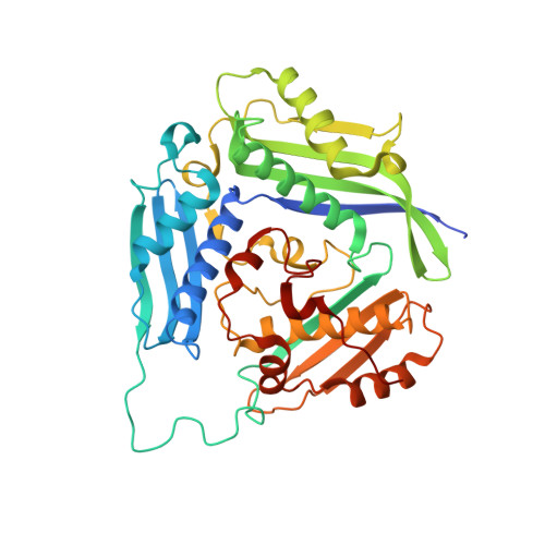
Methionine adenosyltransferase (MAT) deficiency, characterized by isolated persistent hypermethioninemia (IPH), is caused by mutations in the MAT1A gene encoding MATαl, one of the major hepatic enzymes. Most of the associated hypermethioninemic conditions are inherited as autosomal recessive traits; however, dominant inheritance of hypermethioninemia is caused by an Arg264His (R264H) mutation. This mutation has been confirmed in a screening programme of newborns as the most common mutation in babies with IPH. Arg264 makes an inter-subunit salt bridge located at the dimer interface where the active site assembles. Here, it is demonstrated that the R264H mutation results in greatly reduced MAT activity, while retaining its ability to dimerize, indicating that the lower activity arises from alteration at the active site. The first crystallographic structure of the apo form of the wild-type MATαl enzyme is provided, which shows a tetrameric assembly in which two compact dimers combine to form a catalytic tetramer. In contrast, the crystal structure of the MATαl R264H mutant reveals a weaker dimeric assembly, suggesting that the mutation lowers the affinity for dimer-dimer interaction. The formation of a hetero-oligomer with the regulatory MATβV1 subunit or incubation with a quinolone-based compound (SCR0911) results in the near-full recovery of the enzymatic activity of the pathogenic mutation R264H, opening a clear avenue for a therapeutic solution based on chemical interventions that help to correct the defect of the enzyme in its ability to metabolize methionine.
- Molecular Biophysics Group, Institute of Systems, Molecular and Integrative Biology, Faculty of Health and Life Sciences, University of Liverpool, Crown Street, Liverpool L69 7ZB, United Kingdom.
Organizational Affiliation: