6SVS

Crystal Structure of U:A-U-rich RNA triple helix with 11 consecutive base triples


Experimental Data Snapshot

  • Method: X-RAY DIFFRACTION
  • Resolution: 2.50 Å
  • R-Value Free: 0.213 
  • R-Value Work: 0.167 
  • R-Value Observed: 0.169 

wwPDB Validation   3D Report Full Report


Ligand Structure Quality Assessment 


This is version 1.4 of the entry. See complete history


Literature

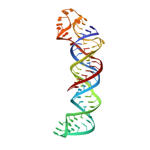
Molecular structure of a U•A-U-rich RNA triple helix with 11 consecutive base triples.

Ruszkowska, A.Ruszkowski, M.Hulewicz, J.P.Dauter, Z.Brown, J.A.

(2020) Nucleic Acids Res 48: 3304-3314

  • DOI: https://doi.org/10.1093/nar/gkz1222
  • Primary Citation of Related Structures:  
    6SVS

  • PubMed Abstract: 

    Three-dimensional structures have been solved for several naturally occurring RNA triple helices, although all are limited to six or fewer consecutive base triples, hindering accurate estimation of global and local structural parameters. We present an X-ray crystal structure of a right-handed, U•A-U-rich RNA triple helix with 11 continuous base triples. Due to helical unwinding, the RNA triple helix spans an average of 12 base triples per turn. The double helix portion of the RNA triple helix is more similar to both the helical and base step structural parameters of A'-RNA rather than A-RNA. Its most striking features are its wide and deep major groove, a smaller inclination angle and all three strands favoring a C3'-endo sugar pucker. Despite the presence of a third strand, the diameter of an RNA triple helix remains nearly identical to those of DNA and RNA double helices. Contrary to our previous modeling predictions, this structure demonstrates that an RNA triple helix is not limited in length to six consecutive base triples and that longer RNA triple helices may exist in nature. Our structure provides a starting point to establish structural parameters of the so-called 'ideal' RNA triple helix, analogous to A-RNA and B-DNA double helices.


  • Organizational Affiliation

    Department of Chemistry and Biochemistry, University of Notre Dame, Notre Dame, IN 46556 USA.


Macromolecules
Find similar nucleic acids by:  (by identity cutoff)  |  3D Structure
Entity ID: 1
MoleculeChains LengthOrganismImage
RNA (79-mer)
A, B
77Homo sapiens
Sequence Annotations
Expand
  • Reference Sequence
Small Molecules
Ligands 4 Unique
IDChains Name / Formula / InChI Key2D Diagram3D Interactions
GTP
Query on GTP

Download Ideal Coordinates CCD File 
H [auth A],
O [auth B]
GUANOSINE-5'-TRIPHOSPHATE
C10 H16 N5 O14 P3
XKMLYUALXHKNFT-UUOKFMHZSA-N
A23
Query on A23

Download Ideal Coordinates CCD File 
I [auth A],
P [auth B]
ADENOSINE-5'-PHOSPHATE-2',3'-CYCLIC PHOSPHATE
C10 H13 N5 O9 P2
HJMBCNJTGVMDOA-KQYNXXCUSA-N
GOL
Query on GOL

Download Ideal Coordinates CCD File 
C [auth A],
J [auth B]
GLYCEROL
C3 H8 O3
PEDCQBHIVMGVHV-UHFFFAOYSA-N
CA
Query on CA

Download Ideal Coordinates CCD File 
D [auth A]
E [auth A]
F [auth A]
G [auth A]
K [auth B]
D [auth A],
E [auth A],
F [auth A],
G [auth A],
K [auth B],
L [auth B],
M [auth B],
N [auth B]
CALCIUM ION
Ca
BHPQYMZQTOCNFJ-UHFFFAOYSA-N
Experimental Data & Validation

Experimental Data

  • Method: X-RAY DIFFRACTION
  • Resolution: 2.50 Å
  • R-Value Free: 0.213 
  • R-Value Work: 0.167 
  • R-Value Observed: 0.169 
  • Space Group: P 1 21 1
Unit Cell:
Length ( Å )Angle ( ˚ )
a = 54.716α = 90
b = 78.114β = 104.2
c = 84.018γ = 90
Software Package:
Software NamePurpose
PHENIXrefinement
PDB_EXTRACTdata extraction
XDSdata reduction
STARANISOdata scaling
PHASERphasing

Structure Validation

View Full Validation Report


Ligand Structure Quality Assessment 


Entry History & Funding Information

Deposition Data


Funding OrganizationLocationGrant Number
National Institutes of Health/National Institute of General Medical Sciences (NIH/NIGMS)United StatesR00GM111430

Revision History  (Full details and data files)

  • Version 1.0: 2020-01-01
    Type: Initial release
  • Version 1.1: 2020-01-29
    Changes: Database references
  • Version 1.2: 2020-04-08
    Changes: Database references
  • Version 1.3: 2022-03-30
    Changes: Author supporting evidence, Database references, Derived calculations
  • Version 1.4: 2024-01-24
    Changes: Data collection, Refinement description