6SSU

Crystal structure of Human Microsomal Glutathione S-Transferase 2 in complex with co-substrate Glutathione


Experimental Data Snapshot

  • Method: X-RAY DIFFRACTION
  • Resolution: 2.50 Å
  • R-Value Free: 0.270 
  • R-Value Work: 0.221 
  • R-Value Observed: 0.224 

wwPDB Validation   3D Report Full Report


Ligand Structure Quality Assessment 


This is version 1.2 of the entry. See complete history


Literature

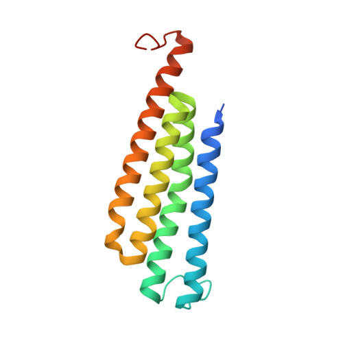
Crystal structures of human MGST2 reveal synchronized conformational changes regulating catalysis.

Thulasingam, M.Orellana, L.Nji, E.Ahmad, S.Rinaldo-Matthis, A.Haeggstrom, J.Z.

(2021) Nat Commun 12: 1728-1728

  • DOI: https://doi.org/10.1038/s41467-021-21924-8
  • Primary Citation of Related Structures:  
    6SSR, 6SSS, 6SSU, 6SSW

  • PubMed Abstract: 

    Microsomal glutathione S-transferase 2 (MGST2) produces leukotriene C 4 , key for intracrine signaling of endoplasmic reticulum (ER) stress, oxidative DNA damage and cell death. MGST2 trimer restricts catalysis to only one out of three active sites at a time, but the molecular basis is unknown. Here, we present crystal structures of human MGST2 combined with biochemical and computational evidence for a concerted mechanism, involving local unfolding coupled to global conformational changes that regulate catalysis. Furthermore, synchronized changes in the biconical central pore modulate the hydrophobicity and control solvent influx to optimize reaction conditions at the active site. These unique mechanistic insights pertain to other, structurally related, drug targets.


  • Organizational Affiliation

    Department of Medical Biochemistry and Biophysics, Division of Chemistry II, Karolinska Institutet, Solnavägen 9, 171 65 Stockholm, Sweden. madhuranayaki.thulasingam@ki.se.


Macromolecules
Find similar proteins by:  (by identity cutoff)  |  3D Structure
Entity ID: 1
MoleculeChains Sequence LengthOrganismDetailsImage
Microsomal glutathione S-transferase 2
A, B, C
153Homo sapiensMutation(s): 0 
Gene Names: MGST2GST2
EC: 2.5.1.18
Membrane Entity: Yes 
UniProt & NIH Common Fund Data Resources
Find proteins for Q99735 (Homo sapiens)
Explore Q99735 
Go to UniProtKB:  Q99735
PHAROS:  Q99735
GTEx:  ENSG00000085871 
Entity Groups  
Sequence Clusters30% Identity50% Identity70% Identity90% Identity95% Identity100% Identity
UniProt GroupQ99735
Sequence Annotations
Expand
  • Reference Sequence
Experimental Data & Validation

Experimental Data

  • Method: X-RAY DIFFRACTION
  • Resolution: 2.50 Å
  • R-Value Free: 0.270 
  • R-Value Work: 0.221 
  • R-Value Observed: 0.224 
  • Space Group: C 2 2 21
Unit Cell:
Length ( Å )Angle ( ˚ )
a = 112.28α = 90
b = 152.37β = 90
c = 71.14γ = 90
Software Package:
Software NamePurpose
PHENIXrefinement
XSCALEdata scaling
PDB_EXTRACTdata extraction
XDSdata reduction
PHENIXphasing

Structure Validation

View Full Validation Report


Ligand Structure Quality Assessment 


Entry History & Funding Information

Deposition Data


Funding OrganizationLocationGrant Number
Swedish Research CouncilSweden10350

Revision History  (Full details and data files)

  • Version 1.0: 2021-02-03
    Type: Initial release
  • Version 1.1: 2021-04-07
    Changes: Data collection, Database references
  • Version 1.2: 2024-01-24
    Changes: Data collection, Database references, Refinement description