6SRE

Crystal Structure of Human Prolidase S202F variant expressed in the presence of chaperones


Experimental Data Snapshot

  • Method: X-RAY DIFFRACTION
  • Resolution: 1.39 Å
  • R-Value Free: 0.172 
  • R-Value Work: 0.150 
  • R-Value Observed: 0.150 

wwPDB Validation   3D Report Full Report


This is version 1.2 of the entry. See complete history


Literature

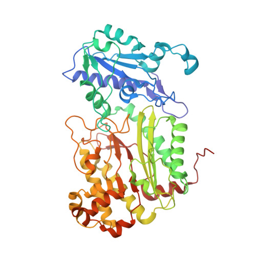
Co-expression with chaperones can affect protein 3D structure as exemplified by loss-of-function variants of human prolidase.

Wator, E.Rutkiewicz, M.Weiss, M.S.Wilk, P.

(2020) FEBS Lett 594: 3045-3056

  • DOI: https://doi.org/10.1002/1873-3468.13877
  • Primary Citation of Related Structures:  
    6SRE, 6SRF, 6SRG

  • PubMed Abstract: 

    Prolidase catalyzes the cleavage of dipeptides containing proline on their C terminus. The reduction in prolidase activity is the cause of a rare disease named 'Prolidase Deficiency'. Local structural disorder was indicated as one of the causes for diminished prolidase activity. Previous studies showed that heat shock proteins can partially recover prolidase activity in vivo. To analyze this mechanism of enzymatic activity rescue, we compared the crystal structures of selected prolidase mutants expressed in the absence and in the presence of chaperones. Our results confirm that protein chaperones facilitate the formation of more ordered structures by their substrate protein. These results also suggest that the protein expression system needs to be considered as an important parameter in structural studies. DATABASES: The reported crystal structures and their associated structure factor amplitudes were deposited in the Protein Data Bank under the accession codes 6SRE, 6SRF, and 6SRG, respectively.


  • Organizational Affiliation

    Macromolecular Crystallography, Helmholtz-Zentrum Berlin für Materialien und Energie, Berlin, Germany.


Macromolecules
Find similar proteins by:  (by identity cutoff)  |  3D Structure
Entity ID: 1
MoleculeChains Sequence LengthOrganismDetailsImage
Xaa-Pro dipeptidase
A, B
484Homo sapiensMutation(s): 1 
Gene Names: PEPDPRD
EC: 3.4.13.9
UniProt & NIH Common Fund Data Resources
Find proteins for P12955 (Homo sapiens)
Explore P12955 
Go to UniProtKB:  P12955
PHAROS:  P12955
GTEx:  ENSG00000124299 
Entity Groups  
Sequence Clusters30% Identity50% Identity70% Identity90% Identity95% Identity100% Identity
UniProt GroupP12955
Sequence Annotations
Expand
  • Reference Sequence
Small Molecules
Ligands 5 Unique
IDChains Name / Formula / InChI Key2D Diagram3D Interactions
PRO
Query on PRO

Download Ideal Coordinates CCD File 
J [auth A],
S [auth B]
PROLINE
C5 H9 N O2
ONIBWKKTOPOVIA-BYPYZUCNSA-N
GOL
Query on GOL

Download Ideal Coordinates CCD File 
D [auth A]
E [auth A]
F [auth A]
G [auth A]
M [auth B]
D [auth A],
E [auth A],
F [auth A],
G [auth A],
M [auth B],
N [auth B],
O [auth B],
P [auth B],
Q [auth B]
GLYCEROL
C3 H8 O3
PEDCQBHIVMGVHV-UHFFFAOYSA-N
GLY
Query on GLY

Download Ideal Coordinates CCD File 
I [auth A],
R [auth B]
GLYCINE
C2 H5 N O2
DHMQDGOQFOQNFH-UHFFFAOYSA-N
MH2
Query on MH2

Download Ideal Coordinates CCD File 
H [auth A],
L [auth B]
MANGANESE ION, 1 HYDROXYL COORDINATED
H Mn O
COIFCHMDWGZDDC-UHFFFAOYSA-M
MN
Query on MN

Download Ideal Coordinates CCD File 
C [auth A],
K [auth B]
MANGANESE (II) ION
Mn
WAEMQWOKJMHJLA-UHFFFAOYSA-N
Experimental Data & Validation

Experimental Data

  • Method: X-RAY DIFFRACTION
  • Resolution: 1.39 Å
  • R-Value Free: 0.172 
  • R-Value Work: 0.150 
  • R-Value Observed: 0.150 
  • Space Group: C 2 2 21
Unit Cell:
Length ( Å )Angle ( ˚ )
a = 103.725α = 90
b = 106.858β = 90
c = 216.323γ = 90
Software Package:
Software NamePurpose
XDSdata reduction
XSCALEdata scaling
PHASERphasing
PHENIXrefinement
PDB_EXTRACTdata extraction

Structure Validation

View Full Validation Report


Entry History 

Deposition Data

Revision History  (Full details and data files)

  • Version 1.0: 2020-07-15
    Type: Initial release
  • Version 1.1: 2020-12-16
    Changes: Database references, Derived calculations
  • Version 1.2: 2024-01-24
    Changes: Data collection, Database references, Refinement description