6SQS

Crystal structure of cat phospho-Ser429 MDM2 RING domain bound to UbcH5B-Ub


Experimental Data Snapshot

  • Method: X-RAY DIFFRACTION
  • Resolution: 1.83 Å
  • R-Value Free: 0.242 
  • R-Value Work: 0.211 

wwPDB Validation   3D Report Full Report


This is version 1.2 of the entry. See complete history


Literature

Structural basis for DNA damage-induced phosphoregulation of MDM2 RING domain.

Magnussen, H.M.Ahmed, S.F.Sibbet, G.J.Hristova, V.A.Nomura, K.Hock, A.K.Archibald, L.J.Jamieson, A.G.Fushman, D.Vousden, K.H.Weissman, A.M.Huang, D.T.

(2020) Nat Commun 11: 2094-2094

  • DOI: https://doi.org/10.1038/s41467-020-15783-y
  • Primary Citation of Related Structures:  
    6SQO, 6SQP, 6SQR, 6SQS

  • PubMed Abstract: 

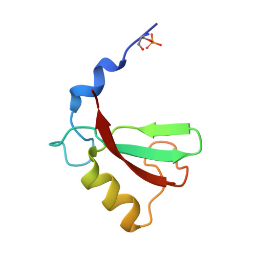
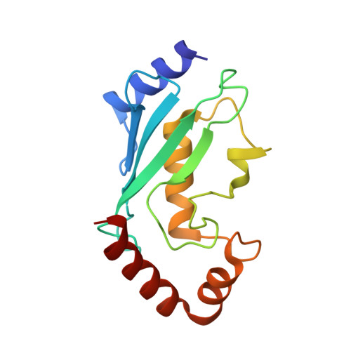
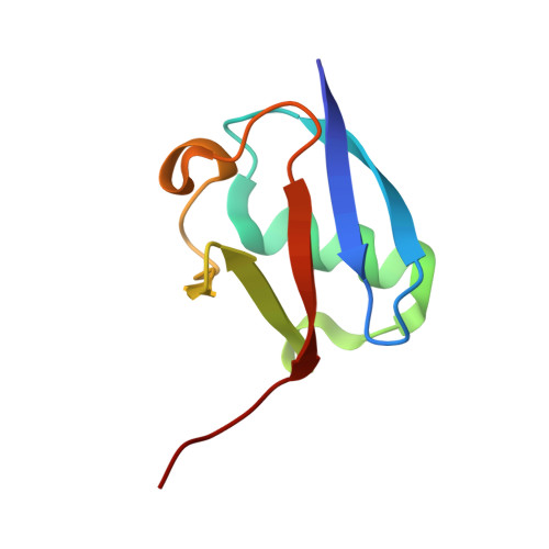
    Phosphorylation of MDM2 by ATM upon DNA damage is an important mechanism for deregulating MDM2, thereby leading to p53 activation. ATM phosphorylates multiple residues near the RING domain of MDM2, but the underlying molecular basis for deregulation remains elusive. Here we show that Ser429 phosphorylation selectively enhances the ubiquitin ligase activity of MDM2 homodimer but not MDM2-MDMX heterodimer. A crystal structure of phospho-Ser429 (pS429)-MDM2 bound to E2-ubiquitin reveals a unique 3 10 -helical feature present in MDM2 homodimer that allows pS429 to stabilize the closed E2-ubiquitin conformation and thereby enhancing ubiquitin transfer. In cells Ser429 phosphorylation increases MDM2 autoubiquitination and degradation upon DNA damage, whereas S429A substitution protects MDM2 from auto-degradation. Our results demonstrate that Ser429 phosphorylation serves as a switch to boost the activity of MDM2 homodimer and promote its self-destruction to enable rapid p53 stabilization and resolve a long-standing controversy surrounding MDM2 auto-degradation in response to DNA damage.


  • Organizational Affiliation

    Cancer Research UK Beatson Institute, Garscube Estate, Switchback Road, Glasgow, G61 1BD, UK.


Macromolecules
Find similar proteins by:  (by identity cutoff)  |  3D Structure
Entity ID: 1
MoleculeChains Sequence LengthOrganismDetailsImage
E3 ubiquitin-protein ligase Mdm2
A, D
64Felis catusMutation(s): 1 
Gene Names: MDM2
EC: 2.3.2.27
UniProt
Find proteins for Q7YRZ8 (Felis catus)
Explore Q7YRZ8 
Go to UniProtKB:  Q7YRZ8
Entity Groups  
Sequence Clusters30% Identity50% Identity70% Identity90% Identity95% Identity100% Identity
UniProt GroupQ7YRZ8
Sequence Annotations
Expand
  • Reference Sequence
Find similar proteins by:  (by identity cutoff)  |  3D Structure
Entity ID: 2
MoleculeChains Sequence LengthOrganismDetailsImage
Ubiquitin-conjugating enzyme E2 D2
B, E
146Homo sapiensMutation(s): 2 
Gene Names: UBE2D2PUBC1UBC4UBC5BUBCH4UBCH5B
EC: 2.3.2.23 (PDB Primary Data), 2.3.2.24 (PDB Primary Data)
UniProt & NIH Common Fund Data Resources
Find proteins for P62837 (Homo sapiens)
Explore P62837 
Go to UniProtKB:  P62837
PHAROS:  P62837
GTEx:  ENSG00000131508 
Entity Groups  
Sequence Clusters30% Identity50% Identity70% Identity90% Identity95% Identity100% Identity
UniProt GroupP62837
Sequence Annotations
Expand
  • Reference Sequence
Find similar proteins by:  (by identity cutoff)  |  3D Structure
Entity ID: 3
MoleculeChains Sequence LengthOrganismDetailsImage
Ubiquitin-40S ribosomal protein S27a
C, F
77Homo sapiensMutation(s): 0 
Gene Names: RPS27A
UniProt & NIH Common Fund Data Resources
Find proteins for P62979 (Homo sapiens)
Explore P62979 
Go to UniProtKB:  P62979
PHAROS:  P62979
GTEx:  ENSG00000143947 
Entity Groups  
Sequence Clusters30% Identity50% Identity70% Identity90% Identity95% Identity100% Identity
UniProt GroupP62979
Sequence Annotations
Expand
  • Reference Sequence
Experimental Data & Validation

Experimental Data

  • Method: X-RAY DIFFRACTION
  • Resolution: 1.83 Å
  • R-Value Free: 0.242 
  • R-Value Work: 0.211 
  • Space Group: P 1
Unit Cell:
Length ( Å )Angle ( ˚ )
a = 54.593α = 66.44
b = 56.438β = 69.44
c = 60.742γ = 89.1
Software Package:
Software NamePurpose
REFMACrefinement
XDSdata reduction
Aimlessdata scaling
PHASERphasing

Structure Validation

View Full Validation Report


Entry History & Funding Information

Deposition Data


Funding OrganizationLocationGrant Number
Cancer Research UKUnited KingdomA23278
European Research CouncilBelgium647849

Revision History  (Full details and data files)

  • Version 1.0: 2020-05-06
    Type: Initial release
  • Version 1.1: 2020-05-13
    Changes: Database references
  • Version 1.2: 2024-01-24
    Changes: Data collection, Database references, Derived calculations, Refinement description