6SAO

Structural and functional characterisation of three novel fungal amylases with enhanced stability and pH tolerance


Experimental Data Snapshot

  • Method: X-RAY DIFFRACTION
  • Resolution: 1.20 Å
  • R-Value Free: 0.134 
  • R-Value Work: 0.110 
  • R-Value Observed: 0.111 

wwPDB Validation   3D Report Full Report


Ligand Structure Quality Assessment 


This is version 2.0 of the entry. See complete history


Literature

Structural and Functional Characterization of Three Novel Fungal Amylases with Enhanced Stability and pH Tolerance.

Roth, C.Moroz, O.V.Turkenburg, J.P.Blagova, E.Waterman, J.Ariza, A.Ming, L.Tianqi, S.Andersen, C.Davies, G.J.Wilson, K.S.

(2019) Int J Mol Sci 20

  • DOI: https://doi.org/10.3390/ijms20194902
  • Primary Citation of Related Structures:  
    6SAO, 6SAU, 6SAV

  • PubMed Abstract: 

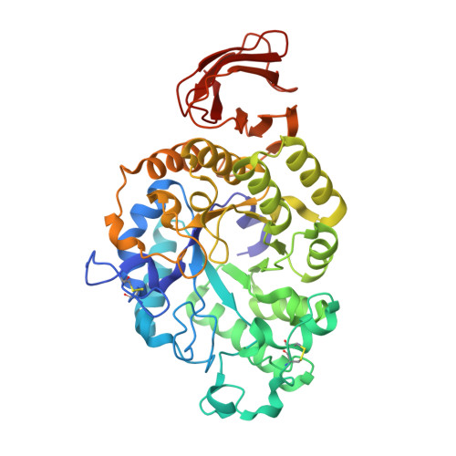
    Amylases are probably the best studied glycoside hydrolases and have a huge biotechnological value for industrial processes on starch. Multiple amylases from fungi and microbes are currently in use. Whereas bacterial amylases are well suited for many industrial processes due to their high stability, fungal amylases are recognized as safe and are preferred in the food industry, although they lack the pH tolerance and stability of their bacterial counterparts. Here, we describe three amylases, two of which have a broad pH spectrum extending to pH 8 and higher stability well suited for a broad set of industrial applications. These enzymes have the characteristic GH13 α-amylase fold with a central (β/α) 8 -domain, an insertion domain with the canonical calcium binding site and a C-terminal β-sandwich domain. The active site was identified based on the binding of the inhibitor acarbose in form of a transglycosylation product, in the amylases from Thamnidium elegans and Cordyceps farinosa . The three amylases have shortened loops flanking the nonreducing end of the substrate binding cleft, creating a more open crevice. Moreover, a potential novel binding site in the C-terminal domain of the Cordyceps enzyme was identified, which might be part of a starch interaction site. In addition, Cordyceps farinosa amylase presented a successful example of using the microseed matrix screening technique to significantly speed-up crystallization.


  • Organizational Affiliation

    York Structural Biology Laboratory, Department of Chemistry, University of York, Heslington, York YO10 5DD, UK. Christian.Roth@mpikg.mpg.de.


Macromolecules
Find similar proteins by:  (by identity cutoff)  |  3D Structure
Entity ID: 1
MoleculeChains Sequence LengthOrganismDetailsImage
amylase439Thamnidium elegansMutation(s): 0 
Entity Groups  
Sequence Clusters30% Identity50% Identity70% Identity90% Identity95% Identity100% Identity
Sequence Annotations
Expand
  • Reference Sequence
Oligosaccharides

Help

Entity ID: 2
MoleculeChains Length2D Diagram Glycosylation3D Interactions
4,6-dideoxy-4-{[(1S,4R,5S,6S)-4,5,6-trihydroxy-3-(hydroxymethyl)cyclohex-2-en-1-yl]amino}-alpha-D-glucopyranose-(1-4)-alpha-D-glucopyranose-(1-4)-4,6-dideoxy-4-{[(1S,4R,5S,6S)-4,5,6-trihydroxy-3-(hydroxymethyl)cyclohex-2-en-1-yl]amino}-alpha-D-glucopyranose-(1-4)-alpha-D-glucopyranose-(1-4)-beta-D-glucopyranose
B
5N/AN/A
Glycosylation Resources
GlyTouCan:  G83366PN
GlyCosmos:  G83366PN
Small Molecules
Ligands 4 Unique
IDChains Name / Formula / InChI Key2D Diagram3D Interactions
NAG
Query on NAG

Download Ideal Coordinates CCD File 
C [auth A]2-acetamido-2-deoxy-beta-D-glucopyranose
C8 H15 N O6
OVRNDRQMDRJTHS-FMDGEEDCSA-N
SO4
Query on SO4

Download Ideal Coordinates CCD File 
D [auth A]
E [auth A]
F [auth A]
I [auth A]
J [auth A]
D [auth A],
E [auth A],
F [auth A],
I [auth A],
J [auth A],
K [auth A]
SULFATE ION
O4 S
QAOWNCQODCNURD-UHFFFAOYSA-L
EDO
Query on EDO

Download Ideal Coordinates CCD File 
G [auth A],
H [auth A]
1,2-ETHANEDIOL
C2 H6 O2
LYCAIKOWRPUZTN-UHFFFAOYSA-N
CA
Query on CA

Download Ideal Coordinates CCD File 
L [auth A]CALCIUM ION
Ca
BHPQYMZQTOCNFJ-UHFFFAOYSA-N
Experimental Data & Validation

Experimental Data

  • Method: X-RAY DIFFRACTION
  • Resolution: 1.20 Å
  • R-Value Free: 0.134 
  • R-Value Work: 0.110 
  • R-Value Observed: 0.111 
  • Space Group: P 21 21 21
Unit Cell:
Length ( Å )Angle ( ˚ )
a = 51.015α = 90
b = 56.633β = 90
c = 166.012γ = 90
Software Package:
Software NamePurpose
REFMACrefinement
PDB_EXTRACTdata extraction
XDSdata reduction
Aimlessdata scaling
MOLREPphasing

Structure Validation

View Full Validation Report


Ligand Structure Quality Assessment 


Entry History 

Deposition Data

Revision History  (Full details and data files)

  • Version 1.0: 2019-10-23
    Type: Initial release
  • Version 1.1: 2019-10-30
    Changes: Data collection, Database references
  • Version 2.0: 2020-07-29
    Type: Remediation
    Reason: Carbohydrate remediation
    Changes: Atomic model, Data collection, Derived calculations, Structure summary