Cycloserine enantiomers are reversible inhibitors of human alanine:glyoxylate aminotransferase: implications for Primary Hyperoxaluria type 1.
Dindo, M., Grottelli, S., Annunziato, G., Giardina, G., Pieroni, M., Pampalone, G., Faccini, A., Cutruzzola, F., Laurino, P., Costantino, G., Cellini, B.(2019) Biochem J 476: 3751-3768
- PubMed: 31794008 Search on PubMed
- DOI: https://doi.org/10.1042/BCJ20190507
- Primary Citation Related Structures:
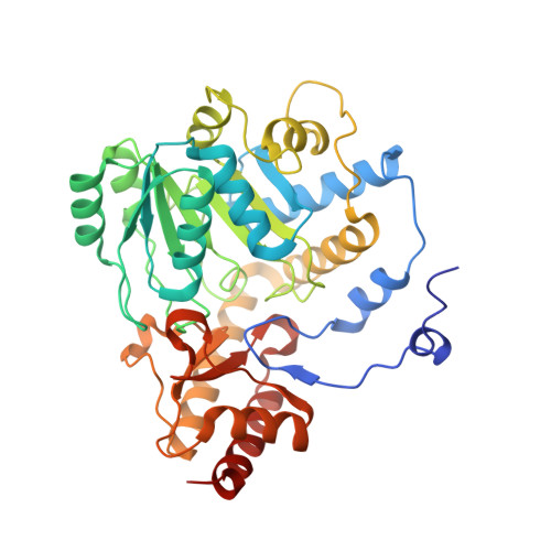
6RV0, 6RV1 - PubMed Abstract:
Peroxisomal alanine:glyoxylate aminotransferase (AGT) is responsible for glyoxylate detoxification in human liver and utilizes pyridoxal 5'-phosphate (PLP) as coenzyme. The deficit of AGT leads to Primary Hyperoxaluria Type I (PH1), a rare disease characterized by calcium oxalate stones deposition in the urinary tract as a consequence of glyoxylate accumulation. Most missense mutations cause AGT misfolding, as in the case of the G41R, which induces aggregation and proteolytic degradation. We have investigated the interaction of wild-type AGT and the pathogenic G41R variant with d-cycloserine (DCS, commercialized as Seromycin), a natural product used as a second-line treatment of multidrug-resistant tuberculosis, and its synthetic enantiomer l-cycloserine (LCS). In contrast with evidences previously reported on other PLP-enzymes, both ligands are AGT reversible inhibitors showing inhibition constants in the micromolar range. While LCS undergoes half-transamination generating a ketimine intermediate and behaves as a classical competitive inhibitor, DCS displays a time-dependent binding mainly generating an oxime intermediate. Using a mammalian cellular model, we found that DCS, but not LCS, is able to promote the correct folding of the G41R variant, as revealed by its increased specific activity and expression as a soluble protein. This effect also translates into an increased glyoxylate detoxification ability of cells expressing the variant upon treatment with DCS. Overall, our findings establish that DCS could play a role as pharmacological chaperone, thus suggesting a new line of intervention against PH1 based on a drug repositioning approach. To a widest extent, this strategy could be applied to other disease-causing mutations leading to AGT misfolding.
- Department of Experimental Medicine, University of Perugia, Perugia, Italy.
Organizational Affiliation: