6RUQ

Structure of GluA2cryst in complex the antagonist ZK200775 and the negative allosteric modulator GYKI53655 at 4.65 A resolution


Experimental Data Snapshot

  • Method: X-RAY DIFFRACTION
  • Resolution: 4.65 Å
  • R-Value Free: 0.291 
  • R-Value Work: 0.242 
  • R-Value Observed: 0.245 

wwPDB Validation   3D Report Full Report


Ligand Structure Quality Assessment 


This is version 2.2 of the entry. See complete history


Literature

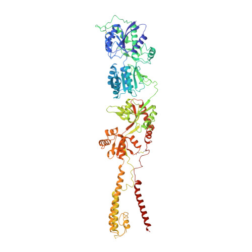
Binding of a negative allosteric modulator and competitive antagonist can occur simultaneously at the ionotropic glutamate receptor GluA2.

Krintel, C.Dorosz, J.Larsen, A.H.Thorsen, T.S.Venskutonyte, R.Mirza, O.Gajhede, M.Boesen, T.Kastrup, J.S.

(2021) FEBS J 288: 995-1007

  • DOI: https://doi.org/10.1111/febs.15455
  • Primary Citation of Related Structures:  
    6RUQ

  • PubMed Abstract: 

    Ionotropic glutamate receptors are ligand-gated ion channels governing neurotransmission in the central nervous system. Three major types of antagonists are known for the AMPA-type receptor GluA2: competitive, noncompetitive (i.e., negative allosteric modulators; NAMs) used for treatment of epilepsy, and uncompetitive antagonists. We here report a 4.65 Å resolution X-ray structure of GluA2, revealing that four molecules of the competitive antagonist ZK200775 and four molecules of the NAM GYKI53655 are capable of binding at the same time. Using negative stain electron microscopy, we show that GYKI53655 alone or ZK200775/GYKI53655 in combination predominantly results in compact receptor forms. The agonist AMPA provides a mixed population of compact and bulgy shapes of GluA2 not impacted by addition of GYKI53655. Taken together, this suggests that the two different mechanisms of antagonism that lead to channel closure are independent and that the distribution between bulgy and compact receptors primarily depends on the ligand bound in the glutamate binding site. DATABASE: The atomic coordinates and structure factors from the crystal structure determination have been deposited in the Protein Data Bank under accession code https://doi.org/10.2210/pdb6RUQ/pdb. The electron microscopy 3D reconstruction volumes have been deposited in EMDB (EMD-4875: Apo; EMD-4920: ZK200775/GYKI53655; EMD-4921: AMPA compact; EMD-4922: AMPA/GYKI53655 bulgy; EMD-4923: GYKI53655; EMD-4924: AMPA bulgy; EMD-4925: AMPA/GYKI53655 compact).


  • Organizational Affiliation

    Research Cluster on Molecular Neuroprotection, Department of Drug Design and Pharmacology, Faculty of Health and Medical Sciences, University of Copenhagen, Denmark.


Macromolecules
Find similar proteins by:  (by identity cutoff)  |  3D Structure
Entity ID: 1
MoleculeChains Sequence LengthOrganismDetailsImage
Glutamate receptor 2
A, B, C, D
823Rattus norvegicusMutation(s): 5 
Gene Names: Gria2Glur2
Membrane Entity: Yes 
UniProt
Find proteins for P19491 (Rattus norvegicus)
Explore P19491 
Go to UniProtKB:  P19491
Entity Groups  
Sequence Clusters30% Identity50% Identity70% Identity90% Identity95% Identity100% Identity
UniProt GroupP19491
Sequence Annotations
Expand
  • Reference Sequence
Oligosaccharides

Help

Entity ID: 2
MoleculeChains Length2D Diagram Glycosylation3D Interactions
beta-D-mannopyranose-(1-4)-2-acetamido-2-deoxy-beta-D-glucopyranose-(1-4)-2-acetamido-2-deoxy-beta-D-glucopyranose
E, G
3N-Glycosylation
Glycosylation Resources
GlyTouCan:  G15407YE
GlyCosmos:  G15407YE
GlyGen:  G15407YE
Entity ID: 3
MoleculeChains Length2D Diagram Glycosylation3D Interactions
beta-D-mannopyranose-(1-3)-beta-D-mannopyranose-(1-4)-2-acetamido-2-deoxy-beta-D-glucopyranose-(1-4)-2-acetamido-2-deoxy-beta-D-glucopyranose
F
4N-Glycosylation
Glycosylation Resources
GlyTouCan:  G31886NL
GlyCosmos:  G31886NL
GlyGen:  G31886NL
Small Molecules
Ligands 2 Unique
IDChains Name / Formula / InChI Key2D Diagram3D Interactions
ZK1 (Subject of Investigation/LOI)
Query on ZK1

Download Ideal Coordinates CCD File 
I [auth A],
K [auth B],
M [auth C],
O [auth D]
{[7-morpholin-4-yl-2,3-dioxo-6-(trifluoromethyl)-3,4-dihydroquinoxalin-1(2H)-yl]methyl}phosphonic acid
C14 H15 F3 N3 O6 P
WZMQMKNCWDCCMT-UHFFFAOYSA-N
GYK (Subject of Investigation/LOI)
Query on GYK

Download Ideal Coordinates CCD File 
H [auth A],
J [auth B],
L [auth C],
N [auth D]
(8R)-5-(4-aminophenyl)-N,8-dimethyl-8,9-dihydro-2H,7H-[1,3]dioxolo[4,5-h][2,3]benzodiazepine-7-carboxamide
C19 H20 N4 O3
SMGACXZFVXKEAX-LLVKDONJSA-N
Experimental Data & Validation

Experimental Data

  • Method: X-RAY DIFFRACTION
  • Resolution: 4.65 Å
  • R-Value Free: 0.291 
  • R-Value Work: 0.242 
  • R-Value Observed: 0.245 
  • Space Group: P 1 21 1
Unit Cell:
Length ( Å )Angle ( ˚ )
a = 91.773α = 90
b = 306.596β = 94.264
c = 107.728γ = 90
Software Package:
Software NamePurpose
MOSFLMdata reduction
Aimlessdata scaling
PHASERphasing
PHENIXrefinement
PDB_EXTRACTdata extraction

Structure Validation

View Full Validation Report


Ligand Structure Quality Assessment 


Entry History & Funding Information

Deposition Data


Funding OrganizationLocationGrant Number
Danish Council for Independent ResearchDenmark--
Other governmentDenmark--
LundbeckfondenDenmark--
Other governmentDenmark--

Revision History  (Full details and data files)

  • Version 1.0: 2020-06-24
    Type: Initial release
  • Version 1.1: 2020-07-01
    Changes: Database references
  • Version 2.0: 2020-07-29
    Type: Remediation
    Reason: Carbohydrate remediation
    Changes: Atomic model, Data collection, Derived calculations, Structure summary
  • Version 2.1: 2021-03-03
    Changes: Database references, Structure summary
  • Version 2.2: 2024-01-24
    Changes: Data collection, Database references, Refinement description