In cellulo crystallization of Trypanosoma brucei IMP dehydrogenase enables the identification of genuine co-factors.
Nass, K., Redecke, L., Perbandt, M., Yefanov, O., Klinge, M., Koopmann, R., Stellato, F., Gabdulkhakov, A., Schonherr, R., Rehders, D., Lahey-Rudolph, J.M., Aquila, A., Barty, A., Basu, S., Doak, R.B., Duden, R., Frank, M., Fromme, R., Kassemeyer, S., Katona, G., Kirian, R., Liu, H., Majoul, I., Martin-Garcia, J.M., Messerschmidt, M., Shoeman, R.L., Weierstall, U., Westenhoff, S., White, T.A., Williams, G.J., Yoon, C.H., Zatsepin, N., Fromme, P., Duszenko, M., Chapman, H.N., Betzel, C.(2020) Nat Commun 11: 620-620
- PubMed: 32001697 Search on PubMedSearch on PubMed Central
- DOI: https://doi.org/10.1038/s41467-020-14484-w
- Primary Citation Related Structures:
6RFU - PubMed Abstract:
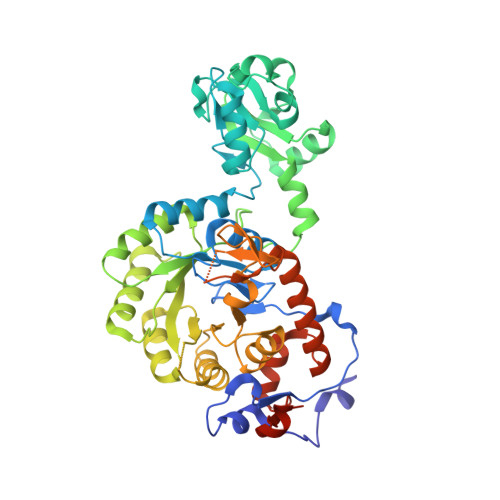
Sleeping sickness is a fatal disease caused by the protozoan parasite Trypanosoma brucei (Tb). Inosine-5'-monophosphate dehydrogenase (IMPDH) has been proposed as a potential drug target, since it maintains the balance between guanylate deoxynucleotide and ribonucleotide levels that is pivotal for the parasite. Here we report the structure of TbIMPDH at room temperature utilizing free-electron laser radiation on crystals grown in living insect cells. The 2.80 Å resolution structure reveals the presence of ATP and GMP at the canonical sites of the Bateman domains, the latter in a so far unknown coordination mode. Consistent with previously reported IMPDH complexes harboring guanosine nucleotides at the second canonical site, TbIMPDH forms a compact oligomer structure, supporting a nucleotide-controlled conformational switch that allosterically modulates the catalytic activity. The oligomeric TbIMPDH structure we present here reveals the potential of in cellulo crystallization to identify genuine allosteric co-factors from a natural reservoir of specific compounds.
- Center for Free-Electron Laser Science (CFEL), Deutsches Elektronen-Synchrotron DESY, Notkestr. 85, 22607, Hamburg, Germany.
Organizational Affiliation: