6QVT

CMP-Sialic acid bound structure of the human wild type Beta-galactoside alpha-2,6-sialyltransferase 1 (ST6Gal1)


Experimental Data Snapshot

  • Method: X-RAY DIFFRACTION
  • Resolution: 1.70 Å
  • R-Value Free: 0.204 
  • R-Value Work: 0.182 
  • R-Value Observed: 0.183 

wwPDB Validation   3D Report Full Report


Ligand Structure Quality Assessment 


This is version 1.5 of the entry. See complete history


Literature

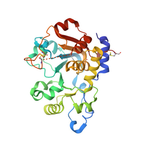
Unliganded and CMP-Neu5Ac bound structures of human alpha-2,6-sialyltransferase ST6Gal I at high resolution.

Harrus, D.Harduin-Lepers, A.Glumoff, T.

(2020) J Struct Biol 212: 107628-107628

  • DOI: https://doi.org/10.1016/j.jsb.2020.107628
  • Primary Citation of Related Structures:  
    6QVS, 6QVT

  • PubMed Abstract: 

    Sialic acid residues found as terminal monosaccharides in various types of glycan chains in cell surface glycoproteins and glycolipids have been identified as important contributors of cell-cell interactions in normal vs. abnormal cellular behavior and are pivotal in diseases such as cancers. In vertebrates, sialic acids are attached to glycan chains by a conserved subset of sialyltransferases with different enzymatic and substrate specificities. ST6Gal I is a sialyltransferase using activated CMP-sialic acids as donor substrates to catalyze the formation of a α2,6-glycosidic bond between the sialic acid residue and the acceptor disaccharide LacNAc. Understanding sialyltransferases at the molecular and structural level shed light into their function. We present here two human ST6Gal I structures, which show for the first time the enzyme in the unliganded state and with the full donor substrate CMP-Neu5Ac bound. Comparison of these structures reveal flexibility of the catalytic loop, since in the unliganded structure Tyr354 adopts a conformation seen also as an alternate conformation in the substrate bound structure. CMP-Neu5Ac is bound with the side chain at C5 of the sugar residue directed outwards at the surface of the protein. Furthermore, the exact binding mode of the sialic acid moiety of the substrate directly involves sialylmotifs L, S and III and positions the sialylmotif VS in the immediate vicinity. We also present a model for the ternary complex of ST6Gal I with both the donor and the acceptor substrates.


  • Organizational Affiliation

    Faculty of Biochemistry and Molecular Medicine, University of Oulu, Aapistie 7A, FI-90220 Oulu, Finland.


Macromolecules
Find similar proteins by:  (by identity cutoff)  |  3D Structure
Entity ID: 1
MoleculeChains Sequence LengthOrganismDetailsImage
Beta-galactoside alpha-2,6-sialyltransferase 1
A, B
275Homo sapiensMutation(s): 0 
Gene Names: ST6GAL1SIAT1
EC: 2.4.99.1
UniProt & NIH Common Fund Data Resources
Find proteins for P15907 (Homo sapiens)
Explore P15907 
Go to UniProtKB:  P15907
PHAROS:  P15907
GTEx:  ENSG00000073849 
Entity Groups  
Sequence Clusters30% Identity50% Identity70% Identity90% Identity95% Identity100% Identity
UniProt GroupP15907
Sequence Annotations
Expand
  • Reference Sequence
Experimental Data & Validation

Experimental Data

  • Method: X-RAY DIFFRACTION
  • Resolution: 1.70 Å
  • R-Value Free: 0.204 
  • R-Value Work: 0.182 
  • R-Value Observed: 0.183 
  • Space Group: C 1 2 1
Unit Cell:
Length ( Å )Angle ( ˚ )
a = 136.661α = 90
b = 93.921β = 94.39
c = 63.718γ = 90
Software Package:
Software NamePurpose
PHENIXrefinement
XDSdata reduction
XSCALEdata scaling
PHASERphasing

Structure Validation

View Full Validation Report


Ligand Structure Quality Assessment 


Entry History & Funding Information

Deposition Data


Funding OrganizationLocationGrant Number
Academy of FinlandFinland285232

Revision History  (Full details and data files)

  • Version 1.0: 2020-06-10
    Type: Initial release
  • Version 1.1: 2020-06-17
    Changes: Database references
  • Version 1.2: 2020-08-19
    Changes: Database references
  • Version 1.3: 2020-09-30
    Changes: Database references
  • Version 1.4: 2020-10-07
    Changes: Database references
  • Version 1.5: 2024-01-24
    Changes: Data collection, Database references, Refinement description