6QP6

Cryo-EM structure of calcium-bound mTMEM16F lipid scramblase in digitonin


Experimental Data Snapshot

  • Method: ELECTRON MICROSCOPY
  • Resolution: 3.20 Å
  • Aggregation State: PARTICLE 
  • Reconstruction Method: SINGLE PARTICLE 

wwPDB Validation   3D Report Full Report


This is version 1.1 of the entry. See complete history


Literature

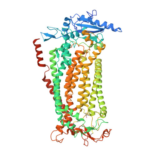
Cryo-EM structures and functional characterization of the murine lipid scramblase TMEM16F.

Alvadia, C.Lim, N.K.Clerico Mosina, V.Oostergetel, G.T.Dutzler, R.Paulino, C.

(2019) Elife 8

  • DOI: https://doi.org/10.7554/eLife.44365
  • Primary Citation of Related Structures:  
    6QP6, 6QPB, 6QPC, 6QPI

  • PubMed Abstract: 

    The lipid scramblase TMEM16F initiates blood coagulation by catalyzing the exposure of phosphatidylserine in platelets. The protein is part of a family of membrane proteins, which encompasses calcium-activated channels for ions and lipids. Here, we reveal features of murine TMEM16F (mTMEM16F) that underlie its function as a lipid scramblase and an ion channel. The cryo-EM data of mTMEM16F in absence and presence of Ca 2+ define the ligand-free closed conformation of the protein and the structure of a Ca 2+ -bound intermediate. Both conformations resemble their counterparts of the scrambling-incompetent anion channel mTMEM16A, yet with distinct differences in the region of ion and lipid permeation. In conjunction with functional data, we demonstrate the relationship between ion conduction and lipid scrambling. Although activated by a common mechanism, both functions appear to be mediated by alternate protein conformations that are at equilibrium in the ligand-bound state.


  • Organizational Affiliation

    Department of Biochemistry, University of Zurich, Zurich, Switzerland.


Macromolecules
Find similar proteins by:  (by identity cutoff)  |  3D Structure
Entity ID: 1
MoleculeChains Sequence LengthOrganismDetailsImage
Anoctamin-6
A, B
911Mus musculusMutation(s): 0 
Gene Names: Ano6Tmem16f
Membrane Entity: Yes 
UniProt & NIH Common Fund Data Resources
Find proteins for Q6P9J9 (Mus musculus)
Explore Q6P9J9 
Go to UniProtKB:  Q6P9J9
IMPC:  MGI:2145890
Entity Groups  
Sequence Clusters30% Identity50% Identity70% Identity90% Identity95% Identity100% Identity
UniProt GroupQ6P9J9
Sequence Annotations
Expand
  • Reference Sequence
Experimental Data & Validation

Experimental Data

  • Method: ELECTRON MICROSCOPY
  • Resolution: 3.20 Å
  • Aggregation State: PARTICLE 
  • Reconstruction Method: SINGLE PARTICLE 
EM Software:
TaskSoftware PackageVersion
RECONSTRUCTIONRELION3.0
MODEL REFINEMENTPHENIX

Structure Validation

View Full Validation Report


Entry History & Funding Information

Deposition Data


Funding OrganizationLocationGrant Number
European Research CouncilSwitzerland339116, AnoBest.
Netherlands Organisation for Scientific ResearchNetherlands740.018.016

Revision History  (Full details and data files)

  • Version 1.0: 2019-03-06
    Type: Initial release
  • Version 1.1: 2019-04-03
    Changes: Data collection, Source and taxonomy