


The structure of GH26C from Muricauda sp. MAR_2010_75
Robb, C.S., Hehemann, J.H.To be published.
Experimental Data Snapshot
Starting Model: experimental
View more details
wwPDB Validation 3D Report Full Report

Entity ID: 1 | |||||
|---|---|---|---|---|---|
| Molecule | Chains | Sequence Length | Organism | Details | Image |
| GH26C | 418 | Allomuricauda sp. MAR_2010_75 | Mutation(s): 0 EC: 3.2.1.78 | 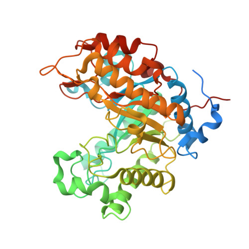 | |
UniProt | |||||
Find proteins for A0ACD6BAS2 (Allomuricauda sp. MAR_2010_75) Explore A0ACD6BAS2 Go to UniProtKB: A0ACD6BAS2 | |||||
Entity Groups | |||||
| Sequence Clusters | 30% Identity50% Identity70% Identity90% Identity95% Identity100% Identity | ||||
| UniProt Group | A0ACD6BAS2 | ||||
Sequence AnnotationsExpand | |||||
Reference Sequence | |||||
| Ligands 1 Unique | |||||
|---|---|---|---|---|---|
| ID | Chains | Name / Formula / InChI Key | 2D Diagram | 3D Interactions | |
| TRS Download:Ideal Coordinates CCD File | C [auth A], D [auth B] | 2-AMINO-2-HYDROXYMETHYL-PROPANE-1,3-DIOL C4 H12 N O3 LENZDBCJOHFCAS-UHFFFAOYSA-O |  | ||
| Length ( Å ) | Angle ( ˚ ) |
|---|---|
| a = 94.119 | α = 90 |
| b = 60.358 | β = 95.92 |
| c = 148.995 | γ = 90 |
| Software Name | Purpose |
|---|---|
| PHENIX | refinement |
| XDS | data reduction |
| Aimless | data scaling |
| PHASER | phasing |
| Funding Organization | Location | Grant Number |
|---|---|---|
| German Research Foundation | Germany | HE 7217/1-1 |


