6Q4B

CDK2 in complex with FragLite13


Experimental Data Snapshot

  • Method: X-RAY DIFFRACTION
  • Resolution: 1.12 Å
  • R-Value Free: 0.203 
  • R-Value Work: 0.173 
  • R-Value Observed: 0.174 

wwPDB Validation   3D Report Full Report


Ligand Structure Quality Assessment 


This is version 1.2 of the entry. See complete history


Literature

FragLites-Minimal, Halogenated Fragments Displaying Pharmacophore Doublets. An Efficient Approach to Druggability Assessment and Hit Generation.

Wood, D.J.Lopez-Fernandez, J.D.Knight, L.E.Al-Khawaldeh, I.Gai, C.Lin, S.Martin, M.P.Miller, D.C.Cano, C.Endicott, J.A.Hardcastle, I.R.Noble, M.E.M.Waring, M.J.

(2019) J Med Chem 62: 3741-3752

  • DOI: https://doi.org/10.1021/acs.jmedchem.9b00304
  • Primary Citation of Related Structures:  
    6Q3B, 6Q3C, 6Q3F, 6Q48, 6Q49, 6Q4A, 6Q4B, 6Q4C, 6Q4D, 6Q4E, 6Q4F, 6Q4G, 6Q4H, 6Q4I, 6Q4J, 6Q4K

  • PubMed Abstract: 

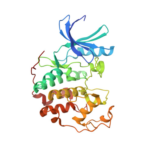
    Identifying ligand binding sites on proteins is a critical step in target-based drug discovery. Current approaches to this require resource-intensive screening of large libraries of lead-like or fragment molecules. Here, we describe an efficient and effective experimental approach to mapping interaction sites using a set of halogenated compounds expressing paired hydrogen-bonding motifs, termed FragLites. The FragLites identify productive drug-like interactions, which are identified sensitively and unambiguously by X-ray crystallography, exploiting the anomalous scattering of the halogen substituent. This mapping of protein interaction surfaces provides an assessment of druggability and can identify efficient start points for the de novo design of hit molecules incorporating the interacting motifs. The approach is illustrated by mapping cyclin-dependent kinase 2, which successfully identifies orthosteric and allosteric sites. The hits were rapidly elaborated to develop efficient lead-like molecules. Hence, the approach provides a new method of identifying ligand sites, assessing tractability and discovering new leads.


  • Organizational Affiliation

    Northern Institute for Cancer Research, Medical School , Newcastle University , Paul O'Gorman Building, Framlington Place , Newcastle upon Tyne NE2 4HH , U.K.


Macromolecules
Find similar proteins by:  (by identity cutoff)  |  3D Structure
Entity ID: 1
MoleculeChains Sequence LengthOrganismDetailsImage
Cyclin-dependent kinase 2306Homo sapiensMutation(s): 0 
Gene Names: CDK2CDKN2
EC: 2.7.11.22
UniProt & NIH Common Fund Data Resources
Find proteins for P24941 (Homo sapiens)
Explore P24941 
Go to UniProtKB:  P24941
PHAROS:  P24941
GTEx:  ENSG00000123374 
Entity Groups  
Sequence Clusters30% Identity50% Identity70% Identity90% Identity95% Identity100% Identity
UniProt GroupP24941
Sequence Annotations
Expand
  • Reference Sequence
Small Molecules
Ligands 1 Unique
IDChains Name / Formula / InChI Key2D Diagram3D Interactions
HHN
Query on HHN

Download Ideal Coordinates CCD File 
B [auth A]5-bromanylpyrimidine
C4 H3 Br N2
GYCPLYCTMDTEPU-UHFFFAOYSA-N
Experimental Data & Validation

Experimental Data

  • Method: X-RAY DIFFRACTION
  • Resolution: 1.12 Å
  • R-Value Free: 0.203 
  • R-Value Work: 0.173 
  • R-Value Observed: 0.174 
  • Space Group: P 21 21 21
Unit Cell:
Length ( Å )Angle ( ˚ )
a = 53.201α = 90
b = 72.191β = 90
c = 72.374γ = 90
Software Package:
Software NamePurpose
REFMACrefinement
xia2data reduction
Aimlessdata scaling
PHASERphasing

Structure Validation

View Full Validation Report


Ligand Structure Quality Assessment 


Entry History & Funding Information

Deposition Data


Funding OrganizationLocationGrant Number
Medical Research Council (United Kingdom)United KingdomMR/N009738/1
Cancer Research UKUnited KingdomC2115/A21421

Revision History  (Full details and data files)

  • Version 1.0: 2019-03-20
    Type: Initial release
  • Version 1.1: 2019-04-24
    Changes: Data collection, Database references
  • Version 1.2: 2024-01-24
    Changes: Data collection, Database references, Refinement description