6Q0Q | pdb_00006q0q

Crystal structure of Human galectin-3 CRD in complex with Methyl 3-O-(1-{3-O-[1-(b-D-galactopyranosyl)-1,2,3-triazol-4-yl]-methyl-b-D-galactopyranosyl}-1,2,3-triazol-4-yl)-methyl-b-D-galactopyranoside


Experimental Data Snapshot

  • Method: X-RAY DIFFRACTION
  • Resolution: 1.99 Å
  • R-Value Free: 
    0.217 (Depositor), 0.218 (DCC) 
  • R-Value Work: 
    0.179 (Depositor), 0.179 (DCC) 
  • R-Value Observed: 
    0.181 (Depositor) 

wwPDB Validation 3D Report Full Report

Validation slider image for 6Q0Q

Ligand Structure Quality Assessment 


This is version 1.2 of the entry. See complete history

Literature

Linear triazole-linked pseudo oligogalactosides as scaffolds for galectin inhibitor development.

Dussouy, C.Kishor, C.Lambert, A.Lamoureux, C.Blanchard, H.Grandjean, C.

(2020) Chem Biol Drug Des 96: 1123-1133

  • DOI: https://doi.org/10.1111/cbdd.13683
  • Primary Citation Related Structures: 
    6Q0Q, 6Q17

  • PubMed Abstract: 

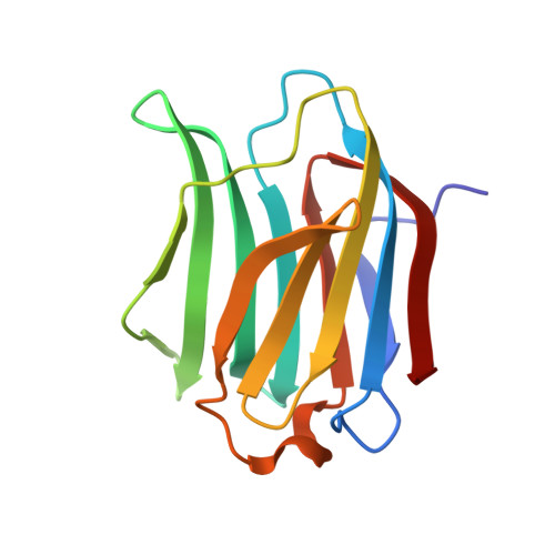
    Galectins play key roles in numerous biological processes. Their mode of action depends on their localization which can be extracellular, cytoplasmic, or nuclear and is partly mediated through interactions with β-galactose containing glycans. Galectins have emerged as novel therapeutic targets notably for the treatment of inflammatory disorders and cancers. This has stimulated the design of carbohydrate-based inhibitors targeting the carbohydrate recognition domains (CRDs) of the galectins. Pursuing this approach, we reasoned that linear oligogalactosides obtained by straightforward iterative click chemistry could mimic poly-lactosamine motifs expressed at eukaryote cell surfaces which the extracellular form of galectin-3, a prominent member of the galectin family, specifically recognizes. Affinities toward galectin-3 consistently increased with the length of the representative oligogalactosides but without reaching that of oligo-lactosamines. Elucidation of the X-ray crystal structures of the galectin-3 CRD in complex with a synthesized di- and tri-galactoside confirmed that the compounds bind within the carbohydrate-binding site. The atomic structures revealed that binding interactions mainly occur with the galactose moiety at the non-reducing end, primarily with subsites C and D of the CRD, differing from oligo-lactosamine which bind more consistently across the whole groove formed by the five subsites (A-E) of the galectin-3 CRD.


  • Organizational Affiliation
    • Unité Fonctionnalité et Ingénierie des Protéines (UFIP), CNRS, UMR 6286, Université de Nantes, Nantes, France.

Macromolecule Content 

  • Total Structure Weight: 16.47 kDa 
  • Atom Count: 1,265 
  • Modeled Residue Count: 139 
  • Deposited Residue Count: 139 
  • Unique protein chains: 1

Macromolecules

Find similar proteins by:|  3D Structure
Entity ID: 1
MoleculeChains  Sequence LengthOrganismDetailsImage
Galectin-3139Homo sapiensMutation(s): 0 
Gene Names: LGALS3MAC2
UniProt & NIH Common Fund Data Resources
Find proteins for P17931 (Homo sapiens)
Explore P17931 
Go to UniProtKB:  P17931
PHAROS:  P17931
GTEx:  ENSG00000131981 
Entity Groups
Sequence Clusters30% Identity50% Identity70% Identity90% Identity95% Identity100% Identity
UniProt GroupP17931
Sequence Annotations
Expand
Reference Sequence

Small Molecules

Ligands 2 Unique
IDChains Name / Formula / InChI Key2D Diagram3D Interactions
P8G
(Subject of Investigation/LOI)

Query on P8G


Download:Ideal Coordinates CCD File
C [auth A](2~{R},3~{R},4~{S},5~{R},6~{R})-2-(hydroxymethyl)-6-[4-[[(2~{R},3~{S},4~{S},5~{R},6~{R})-2-(hydroxymethyl)-6-[4-[[(2~{R},3~{S},4~{S},5~{R},6~{R})-2-(hydroxymethyl)-6-methoxy-3,5-bis(oxidanyl)oxan-4-yl]oxymethyl]-1,2,3-triazol-1-yl]-3,5-bis(oxidanyl)oxan-4-yl]oxymethyl]-1,2,3-triazol-1-yl]oxane-3,4,5-triol
C25 H40 N6 O16
ZQXDLGAORGHMLS-LZGLEUEQSA-N
CL

Query on CL


Download:Ideal Coordinates CCD File
B [auth A]CHLORIDE ION
Cl
VEXZGXHMUGYJMC-UHFFFAOYSA-M

Experimental Data & Validation

Experimental Data

  • Method: X-RAY DIFFRACTION
  • Resolution: 1.99 Å
  • R-Value Free:  0.217 (Depositor), 0.218 (DCC) 
  • R-Value Work:  0.179 (Depositor), 0.179 (DCC) 
  • R-Value Observed: 0.181 (Depositor) 
Space Group: P 21 21 21
Unit Cell:
Length ( Å )Angle ( ˚ )
a = 35.95α = 90
b = 57.86β = 90
c = 62.34γ = 90
Software Package:
Software NamePurpose
PHENIXrefinement
MOSFLMdata reduction
SCALAdata scaling

Structure Validation

View Full Validation Report


Ligand Structure Quality Assessment 


Entry History 

& Funding Information

Deposition Data


Funding OrganizationLocationGrant Number
Other governmentAustraliaCancer Council Queensland Grant ID180845

Revision History  (Full details and data files)

  • Version 1.0: 2020-04-29
    Type: Initial release
  • Version 1.1: 2020-12-23
    Changes: Database references
  • Version 1.2: 2024-03-13
    Changes: Data collection, Database references