6PV9 | pdb_00006pv9

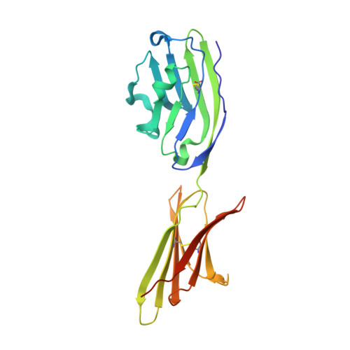
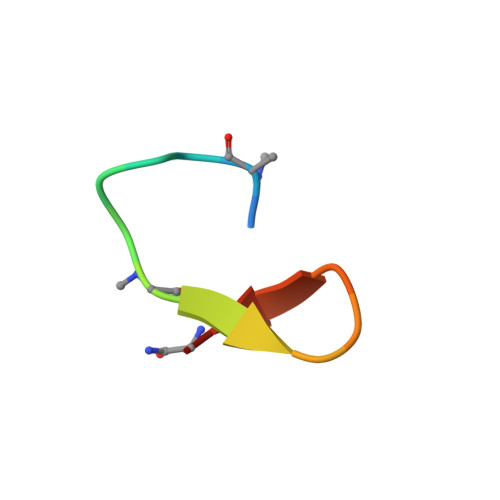
Human PD-L1 bound to a macrocyclic peptide which blocks the PD-1/PD-L1 interaction


Experimental Data Snapshot

  • Method: X-RAY DIFFRACTION
  • Resolution: 2.00 Å
  • R-Value Free: 
    0.226 (Depositor), 0.226 (DCC) 
  • R-Value Work: 
    0.191 (Depositor), 0.191 (DCC) 
  • R-Value Observed: 
    0.194 (Depositor) 

Starting Model: other
View more details

wwPDB Validation 3D Report Full Report

Validation slider image for 6PV9

This is version 1.2 of the entry. See complete history. 

Literature

Protein Footprinting and X-ray Crystallography Reveal the Interaction of PD-L1 and a Macrocyclic Peptide.

Niu, B., Appleby, T.C., Wang, R., Morar, M., Voight, J., Villasenor, A.G., Clancy, S., Wise, S., Belzile, J.P., Papalia, G., Wong, M., Brendza, K.M., Lad, L., Gross, M.L.

(2020) Biochemistry 59: 541-551

  • DOI: https://doi.org/10.1021/acs.biochem.9b00822
  • Primary Citation Related Structures: 
    6PV9

  • PubMed Abstract: 

    Blocking interactions between PD-1 and PD-L1 opens a new era of cancer treatment involving immunity modulation. Although most immunotherapies use monoclonal antibodies, small-molecule inhibitors offer advantages. To facilitate development of small-molecule therapeutics, we implemented a rapid approach to characterize the binding interfaces of small-molecule inhibitors with PD-L1. We determined its interaction with a synthetic macrocyclic peptide by using two mass spectrometry-based approaches, hydrogen-deuterium exchange and fast photochemical oxidation of proteins (FPOP), and corroborated the findings with our X-ray structure of the PD-L1/macrocycle complex. Although all three approaches show that the macrocycle binds directly to PD-L1 over the regions of residues 46-87 and 114-125, the two protein footprinting approaches show additional binding at the N-terminus of PD-L1, and FPOP reveals some critical binding residues. The outcomes not only show the binding regions but also demonstrate the utility of MS-based footprinting in probing protein/ligand inhibitory interactions in cancer immunotherapy.


  • Organizational Affiliation: 
    • Department of Chemistry , Washington University in St. Louis , St. Louis , Missouri 63130 , United States.

Macromolecule Content 

  • Total Structure Weight: 27.3 kDa 
  • Atom Count: 1,902 
  • Modeled Residue Count: 222 
  • Deposited Residue Count: 239 
  • Unique protein chains: 2

Macromolecules

Find similar proteins by:|  3D Structure
Entity ID: 1
MoleculeChains  Sequence LengthOrganismDetailsImage
Programmed cell death 1 ligand 1222Homo sapiensMutation(s): 0 
Gene Names: CD274, B7H1, PDCD1L1, PDCD1LG1, PDL1
UniProt & NIH Common Fund Data Resources
Find proteins for Q9NZQ7 (Homo sapiens)
Explore Q9NZQ7 
Go to UniProtKB:  Q9NZQ7
PHAROS:  Q9NZQ7
GTEx:  ENSG00000120217 
Entity Groups
Sequence Clusters30% Identity50% Identity70% Identity90% Identity95% Identity100% Identity
UniProt GroupQ9NZQ7
Sequence Annotations
Expand
Reference Sequence
Find similar proteins by:  Sequence   |   3D Structure  
Entity ID: 2
MoleculeChains  Sequence LengthOrganismDetailsImage
macrocyclic peptide17synthetic constructMutation(s): 0 
Entity Groups
Sequence Clusters30% Identity50% Identity70% Identity90% Identity95% Identity100% Identity
Sequence Annotations
Expand
Reference Sequence

Small Molecules

Non-standard Monomers  4 Unique
IDChains TypeFormula2D Diagram
9KK
Query on 9KK
L-PEPTIDE LINKINGC7 H15 N O2
DCY
Query on DCY
D-PEPTIDE LINKINGC3 H7 N O2 S
MAA
Query on MAA
L-PEPTIDE LINKINGC4 H9 N O2
SAR
Query on SAR
PEPTIDE LINKINGC3 H7 N O2

Experimental Data & Validation

Experimental Data

  • Method: X-RAY DIFFRACTION
  • Resolution: 2.00 Å
  • R-Value Free:  0.226 (Depositor), 0.226 (DCC) 
  • R-Value Work:  0.191 (Depositor), 0.191 (DCC) 
  • R-Value Observed: 0.194 (Depositor) 
Space Group: P 64
Unit Cell:
Length ( Å )Angle ( ˚ )
a = 114.949α = 90
b = 114.949β = 90
c = 33.778γ = 120
Software Package:
Software NamePurpose
HKL-2000data reduction
SCALEPACKdata scaling
PHENIXrefinement
PDB_EXTRACTdata extraction
PHENIXphasing

Structure Validation

View Full Validation Report


Entry History 

& Funding Information

Deposition Data


Funding OrganizationLocationGrant Number
National Institutes of Health/National Institute of General Medical Sciences (NIH/NIGMS)United States2P41GM103422

Revision History  (Full details and data files)

  • Version 1.0: 2020-01-01
    Type: Initial release
  • Version 1.1: 2020-02-19
    Changes: Database references
  • Version 1.2: 2024-04-03
    Changes: Data collection, Database references, Derived calculations, Refinement description