6PV9 | pdb_00006pv9

Human PD-L1 bound to a macrocyclic peptide which blocks the PD-1/PD-L1 interaction


Experimental Data Snapshot

  • Method: X-RAY DIFFRACTION
  • Resolution: 2.00 Å
  • R-Value Free: 
    0.226 (Depositor), 0.226 (DCC) 
  • R-Value Work: 
    0.191 (Depositor), 0.191 (DCC) 
  • R-Value Observed: 
    0.194 (Depositor) 

Starting Model: other
View more details

wwPDB Validation 3D Report Full Report

Validation slider image for 6PV9

This is version 1.2 of the entry. See complete history

Literature

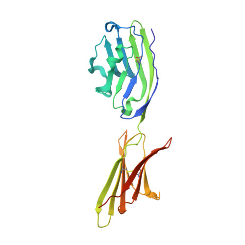
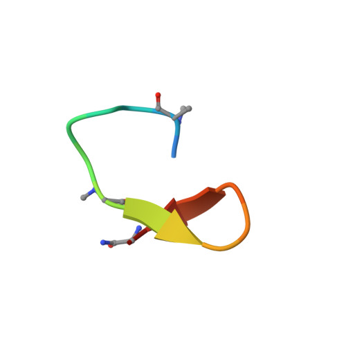
Protein Footprinting and X-ray Crystallography Reveal the Interaction of PD-L1 and a Macrocyclic Peptide.

Niu, B.Appleby, T.C.Wang, R.Morar, M.Voight, J.Villasenor, A.G.Clancy, S.Wise, S.Belzile, J.P.Papalia, G.Wong, M.Brendza, K.M.Lad, L.Gross, M.L.

(2020) Biochemistry 59: 541-551

  • DOI: https://doi.org/10.1021/acs.biochem.9b00822
  • Primary Citation Related Structures: 
    6PV9

  • PubMed Abstract: 

    Blocking interactions between PD-1 and PD-L1 opens a new era of cancer treatment involving immunity modulation. Although most immunotherapies use monoclonal antibodies, small-molecule inhibitors offer advantages. To facilitate development of small-molecule therapeutics, we implemented a rapid approach to characterize the binding interfaces of small-molecule inhibitors with PD-L1. We determined its interaction with a synthetic macrocyclic peptide by using two mass spectrometry-based approaches, hydrogen-deuterium exchange and fast photochemical oxidation of proteins (FPOP), and corroborated the findings with our X-ray structure of the PD-L1/macrocycle complex. Although all three approaches show that the macrocycle binds directly to PD-L1 over the regions of residues 46-87 and 114-125, the two protein footprinting approaches show additional binding at the N-terminus of PD-L1, and FPOP reveals some critical binding residues. The outcomes not only show the binding regions but also demonstrate the utility of MS-based footprinting in probing protein/ligand inhibitory interactions in cancer immunotherapy.


  • Organizational Affiliation
    • Department of Chemistry , Washington University in St. Louis , St. Louis , Missouri 63130 , United States.

Macromolecule Content 

  • Total Structure Weight: 27.3 kDa 
  • Atom Count: 1,902 
  • Modeled Residue Count: 222 
  • Deposited Residue Count: 239 
  • Unique protein chains: 2

Macromolecules

Find similar proteins by:|  3D Structure
Entity ID: 1
MoleculeChains  Sequence LengthOrganismDetailsImage
Programmed cell death 1 ligand 1222Homo sapiensMutation(s): 0 
Gene Names: CD274B7H1PDCD1L1PDCD1LG1PDL1
UniProt & NIH Common Fund Data Resources
Find proteins for Q9NZQ7 (Homo sapiens)
Explore Q9NZQ7 
Go to UniProtKB:  Q9NZQ7
PHAROS:  Q9NZQ7
GTEx:  ENSG00000120217 
Entity Groups
Sequence Clusters30% Identity50% Identity70% Identity90% Identity95% Identity100% Identity
UniProt GroupQ9NZQ7
Sequence Annotations
Expand
Reference Sequence
Find similar proteins by:  Sequence   |   3D Structure  
Entity ID: 2
MoleculeChains  Sequence LengthOrganismDetailsImage
macrocyclic peptide17synthetic constructMutation(s): 0 

Small Molecules

Modified Residues  2 Unique
IDChains TypeFormula2D DiagramParent
9KK
Query on 9KK
B
L-PEPTIDE LINKINGC7 H15 N O2

--

DCY
Query on DCY
B
D-PEPTIDE LINKINGC3 H7 N O2 S

--

MAA
Query on MAA
B
L-PEPTIDE LINKINGC4 H9 N O2ALA
SAR
Query on SAR
B
PEPTIDE LINKINGC3 H7 N O2GLY

Experimental Data & Validation

Experimental Data

  • Method: X-RAY DIFFRACTION
  • Resolution: 2.00 Å
  • R-Value Free:  0.226 (Depositor), 0.226 (DCC) 
  • R-Value Work:  0.191 (Depositor), 0.191 (DCC) 
  • R-Value Observed: 0.194 (Depositor) 
Space Group: P 64
Unit Cell:
Length ( Å )Angle ( ˚ )
a = 114.949α = 90
b = 114.949β = 90
c = 33.778γ = 120
Software Package:
Software NamePurpose
HKL-2000data reduction
SCALEPACKdata scaling
PHENIXrefinement
PDB_EXTRACTdata extraction
PHENIXphasing

Structure Validation

View Full Validation Report


Entry History 

& Funding Information

Deposition Data


Funding OrganizationLocationGrant Number
National Institutes of Health/National Institute of General Medical Sciences (NIH/NIGMS)United States2P41GM103422

Revision History  (Full details and data files)

  • Version 1.0: 2020-01-01
    Type: Initial release
  • Version 1.1: 2020-02-19
    Changes: Database references
  • Version 1.2: 2024-04-03
    Changes: Data collection, Database references, Derived calculations, Refinement description