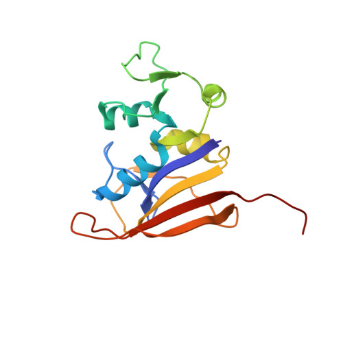
6PR9

S. aureus dihydrofolate reductase co-crystallized with 1-ethylpropyl-dihydropthalazine inhibitor and NADP(H)


Experimental Data Snapshot

  • Method: X-RAY DIFFRACTION
  • Resolution: 2.01 Å
  • R-Value Free: 0.219 
  • R-Value Work: 0.172 
  • R-Value Observed: 0.175 

wwPDB Validation   3D Report Full Report


This is version 2.0 of the entry. See complete history


Literature

Inhibitor design to target a unique feature in the folate pocket of Staphylococcus aureus dihydrofolate reductase.

Muddala, N.P.White, J.C.Nammalwar, B.Pratt, I.Thomas, L.M.Bunce, R.A.Berlin, K.D.Bourne, C.R.

(2020) Eur J Med Chem 200: 112412-112412

  • DOI: https://doi.org/10.1016/j.ejmech.2020.112412
  • Primary Citation of Related Structures:  
    6PR6, 6PR7, 6PR8, 6PR9, 6PRA, 6PRB, 6PRD

  • PubMed Abstract: 

    Staphylococcus aureus (Sa) is a serious concern due to increasing resistance to antibiotics. The bacterial dihydrofolate reductase enzyme is effectively inhibited by trimethoprim, a compound with antibacterial activity. Previously, we reported a trimethoprim derivative containing an acryloyl linker and a dihydophthalazine moiety demonstrating increased potency against S. aureus. We have expanded this series and assessed in vitro enzyme inhibition (K i ) and whole cell growth inhibition properties (MIC). Modifications were focused at a chiral carbon within the phthalazine heterocycle, as well as simultaneous modification at positions on the dihydrophthalazine. MIC values increased from 0.0626-0.5 μg/mL into the 0.5-1 μg/mL range when the edge positions were modified with either methyl or methoxy groups. Changes at the chiral carbon affected K i measurements but with little impact on MIC values. Our structural data revealed accommodation of predominantly the S-enantiomer of the inhibitors within the folate-binding pocket. Longer modifications at the chiral carbon, such as p-methylbenzyl, protrude from the pocket into solvent and result in poorer K i values, as do modifications with greater torsional freedom, such as 1-ethylpropyl. The most efficacious K i was 0.7 ± 0.3 nM, obtained with a cyclopropyl derivative containing dimethoxy modifications at the dihydrophthalazine edge. The co-crystal structure revealed an alternative placement of the phthalazine moiety into a shallow surface at the edge of the site that can accommodate either enantiomer of the inhibitor. The current design, therefore, highlights how to engineer specific placement of the inhibitor within this alternative pocket, which in turn maximizes the enzyme inhibitory properties of racemic mixtures.


  • Organizational Affiliation

    Department of Chemistry, Oklahoma State University, 107 Physical Sciences I, Stillwater, OK, 74078, USA.


Macromolecules
Find similar proteins by:  (by identity cutoff)  |  3D Structure
Entity ID: 1
MoleculeChains Sequence LengthOrganismDetailsImage
Dihydrofolate reductase162Staphylococcus aureusMutation(s): 0 
Gene Names: folA
EC: 1.5.1.3
UniProt
Find proteins for P0A017 (Staphylococcus aureus)
Explore P0A017 
Go to UniProtKB:  P0A017
Entity Groups  
Sequence Clusters30% Identity50% Identity70% Identity90% Identity95% Identity100% Identity
UniProt GroupP0A017
Sequence Annotations
Expand
  • Reference Sequence
Small Molecules
Ligands 3 Unique
IDChains Name / Formula / InChI Key2D Diagram3D Interactions
NAP
Query on NAP

Download Ideal Coordinates CCD File 
B [auth A]NADP NICOTINAMIDE-ADENINE-DINUCLEOTIDE PHOSPHATE
C21 H28 N7 O17 P3
XJLXINKUBYWONI-NNYOXOHSSA-N
OWV (Subject of Investigation/LOI)
Query on OWV

Download Ideal Coordinates CCD File 
C [auth A](2E)-3-{5-[(2,4-diaminopyrimidin-5-yl)methyl]-2,3-dimethoxyphenyl}-1-[(1S)-1-(pentan-3-yl)-3,4-dihydrophthalazin-2(1H)-yl]prop-2-en-1-one
C29 H36 N6 O3
NVYBVAIPWQNGSG-GIPLCNRYSA-N
GOL
Query on GOL

Download Ideal Coordinates CCD File 
D [auth A],
E [auth A]
GLYCEROL
C3 H8 O3
PEDCQBHIVMGVHV-UHFFFAOYSA-N
Binding Affinity Annotations 
IDSourceBinding Affinity
OWV Binding MOAD:  6PR9 Ki: 2.6 (nM) from 1 assay(s)
Experimental Data & Validation

Experimental Data

  • Method: X-RAY DIFFRACTION
  • Resolution: 2.01 Å
  • R-Value Free: 0.219 
  • R-Value Work: 0.172 
  • R-Value Observed: 0.175 
  • Space Group: P 61 2 2
Unit Cell:
Length ( Å )Angle ( ˚ )
a = 78.98α = 90
b = 78.98β = 90
c = 107.33γ = 120
Software Package:
Software NamePurpose
PHENIXrefinement
CrystalCleardata collection
PHASERphasing
MOSFLMdata reduction
Aimlessdata scaling
PHENIXphasing

Structure Validation

View Full Validation Report


Entry History & Funding Information

Deposition Data


Funding OrganizationLocationGrant Number
National Institutes of Health/National Institute Of Allergy and Infectious Diseases (NIH/NIAID)United StatesR01-AI090685-01

Revision History  (Full details and data files)

  • Version 1.0: 2020-06-17
    Type: Initial release
  • Version 2.0: 2023-10-11
    Changes: Data collection, Database references, Non-polymer description, Refinement description