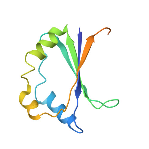
Structure of aMycobacterium tuberculosisHeme-Degrading Protein, MhuD, Variant in Complex with Its Product.
Chao, A., Burley, K.H., Sieminski, P.J., de Miranda, R., Chen, X., Mobley, D.L., Goulding, C.W.(2019) Biochemistry 58: 4610-4620
- PubMed: 31638374 Search on PubMedSearch on PubMed Central
- DOI: https://doi.org/10.1021/acs.biochem.9b00726
- Primary Citation Related Structures:
6PLE - PubMed Abstract:
Mycobacterium tuberculosis (Mtb), the causative agent of tuberculosis, requires iron for survival. In Mtb, MhuD is the cytosolic protein that degrades imported heme. MhuD is distinct, in both sequence and structure, from canonical heme oxygenases (HOs) but homologous with IsdG-type proteins. Canonical HO is found mainly in eukaryotes, while IsdG-type proteins are predominantly found in prokaryotes, including pathogens. While there are several published structures of MhuD and other IsdG-type proteins in complex with the heme substrate, no structures of IsdG-type proteins in complex with a product have been reported, unlike the case for HOs. We recently showed that the Mtb variant MhuD-R26S produces biliverdin IXα (αBV) rather than the wild-type mycobilin isomers. Given that mycobilin and other IsdG-type protein products like staphylobilin are difficult to isolate in quantities sufficient for structure determination, here we use the MhuD-R26S variant and its product αBV as a proxy to study the IsdG-type protein-product complex. First, we show that αBV has a nanomolar affinity for MhuD and the R26S variant. Second, we determined the MhuD-R26S-αBV complex structure to 2.5 Å, which reveals two notable features: (1) two αBV molecules bound per active site and (2) a novel α-helix (α3) that was not observed in previous MhuD-heme structures. Finally, through molecular dynamics simulations, we show that α3 is stable with the proximal αBV alone. MhuD's high affinity for the product and the observed structural and electrostatic changes that accompany substrate turnover suggest that there may be an unidentified class of proteins that are responsible for the extraction of products from MhuD and other IsdG-type proteins.