Substrate-Triggered Formation of a Peroxo-Fe2(III/III) Intermediate during Fatty Acid Decarboxylation by UndA.
Zhang, B., Rajakovich, L.J., Van Cura, D., Blaesi, E.J., Mitchell, A.J., Tysoe, C.R., Zhu, X., Streit, B.R., Rui, Z., Zhang, W., Boal, A.K., Krebs, C., Bollinger Jr., J.M.(2019) J Am Chem Soc 141: 14510-14514
- PubMed: 31487162 Search on PubMedSearch on PubMed Central
- DOI: https://doi.org/10.1021/jacs.9b06093
- Primary Citation Related Structures:
6P5Q - PubMed Abstract:
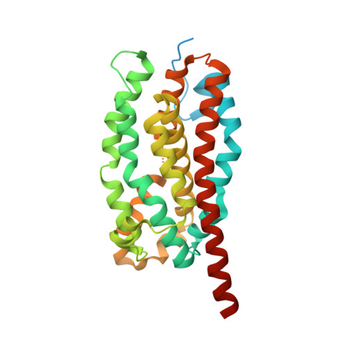
The iron-dependent oxidase UndA cleaves one C3-H bond and the C1-C2 bond of dodecanoic acid to produce 1-undecene and CO 2 . A published X-ray crystal structure showed that UndA has a heme-oxygenase-like fold, thus associating it with a structural superfamily that includes known and postulated non-heme diiron proteins, but revealed only a single iron ion in the active site. Mechanisms proposed for initiation of decarboxylation by cleavage of the C3-H bond using a monoiron cofactor to activate O 2 necessarily invoked unusual or potentially unfeasible steps. Here we present spectroscopic, crystallographic, and biochemical evidence that the cofactor of Pseudomonas fluorescens Pf-5 UndA is actually a diiron cluster and show that binding of the substrate triggers rapid addition of O 2 to the Fe 2 (II/II) cofactor to produce a transient peroxo-Fe 2 (III/III) intermediate. The observations of a diiron cofactor and substrate-triggered formation of a peroxo-Fe 2 (III/III) intermediate suggest a small set of possible mechanisms for O 2 , C3-H and C1-C2 activation by UndA; these routes obviate the problematic steps of the earlier hypotheses that invoked a single iron.
- Department of Chemical and Biomolecular Engineering , University of California Berkeley , Berkeley , California 94720 , United States.
Organizational Affiliation: