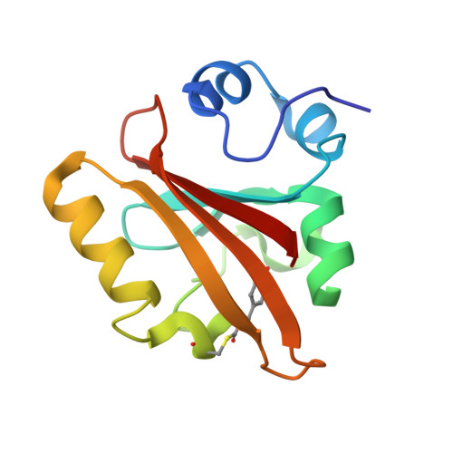
Time-resolved serial femtosecond crystallography at the European XFEL.
Pandey, S., Bean, R., Sato, T., Poudyal, I., Bielecki, J., Cruz Villarreal, J., Yefanov, O., Mariani, V., White, T.A., Kupitz, C., Hunter, M., Abdellatif, M.H., Bajt, S., Bondar, V., Echelmeier, A., Doppler, D., Emons, M., Frank, M., Fromme, R., Gevorkov, Y., Giovanetti, G., Jiang, M., Kim, D., Kim, Y., Kirkwood, H., Klimovskaia, A., Knoska, J., Koua, F.H.M., Letrun, R., Lisova, S., Maia, L., Mazalova, V., Meza, D., Michelat, T., Ourmazd, A., Palmer, G., Ramilli, M., Schubert, R., Schwander, P., Silenzi, A., Sztuk-Dambietz, J., Tolstikova, A., Chapman, H.N., Ros, A., Barty, A., Fromme, P., Mancuso, A.P., Schmidt, M.(2020) Nat Methods 17: 73-78
- PubMed: 31740816
- DOI: https://doi.org/10.1038/s41592-019-0628-z
- Primary Citation of Related Structures:
6P4I, 6P5D, 6P5E, 6P5F, 6P5G - PubMed Abstract:
The European XFEL (EuXFEL) is a 3.4-km long X-ray source, which produces femtosecond, ultrabrilliant and spatially coherent X-ray pulses at megahertz (MHz) repetition rates. This X-ray source has been designed to enable the observation of ultrafast processes with near-atomic spatial resolution. Time-resolved crystallographic investigations on biological macromolecules belong to an important class of experiments that explore fundamental and functional structural displacements in these molecules. Due to the unusual MHz X-ray pulse structure at the EuXFEL, these experiments are challenging. Here, we demonstrate how a biological reaction can be followed on ultrafast timescales at the EuXFEL. We investigate the picosecond time range in the photocycle of photoactive yellow protein (PYP) with MHz X-ray pulse rates. We show that difference electron density maps of excellent quality can be obtained. The results connect the previously explored femtosecond PYP dynamics to timescales accessible at synchrotrons. This opens the door to a wide range of time-resolved studies at the EuXFEL.
- Physics Department, University of Wisconsin-Milwaukee, Milwaukee, WI, USA.
Organizational Affiliation:


