6P2D | pdb_00006p2d

Structure of mouse ketohexokinase-C in complex with fructose and ADP


Experimental Data Snapshot

  • Method: X-RAY DIFFRACTION
  • Resolution: 1.79 Å
  • R-Value Free: 
    0.220 (Depositor), 0.217 (DCC) 
  • R-Value Work: 
    0.170 (Depositor), 0.170 (DCC) 

Starting Model: experimental
View more details

wwPDB Validation 3D Report Full Report

Validation slider image for 6P2D

Ligand Structure Quality Assessment 


This is version 2.2 of the entry. See complete history

Literature

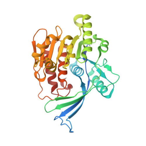
Michaelis-like complex of mouse ketohexokinase isoform C.

Gasper, W.C.Gardner, S.Ross, A.Oppelt, S.A.Allen, K.N.Tolan, D.R.

(2024) Acta Crystallogr D Struct Biol 80: 377-385

  • DOI: https://doi.org/10.1107/S2059798324003723
  • Primary Citation Related Structures: 
    6P2D

  • PubMed Abstract: 

    Over the past forty years there has been a drastic increase in fructose-related diseases, including obesity, heart disease and diabetes. Ketohexokinase (KHK), the first enzyme in the liver fructolysis pathway, catalyzes the ATP-dependent phosphorylation of fructose to fructose 1-phosphate. Understanding the role of KHK in disease-related processes is crucial for the management and prevention of this growing epidemic. Molecular insight into the structure-function relationship in ligand binding and catalysis by KHK is needed for the design of therapeutic inhibitory ligands. Ketohexokinase has two isoforms: ketohexokinase A (KHK-A) is produced ubiquitously at low levels, whereas ketohexokinase C (KHK-C) is found at much higher levels, specifically in the liver, kidneys and intestines. Structures of the unliganded and liganded human isoforms KHK-A and KHK-C are known, as well as structures of unliganded and inhibitor-bound mouse KHK-C (mKHK-C), which shares 90% sequence identity with human KHK-C. Here, a high-resolution X-ray crystal structure of mKHK-C refined to 1.79 Å resolution is presented. The structure was determined in a complex with both the substrate fructose and the product of catalysis, ADP, providing a view of the Michaelis-like complex of the mouse ortholog. Comparison to unliganded structures suggests that KHK undergoes a conformational change upon binding of substrates that places the enzyme in a catalytically competent form in which the β-sheet domain from one subunit rotates by 16.2°, acting as a lid for the opposing active site. Similar kinetic parameters were calculated for the mouse and human enzymes and indicate that mice may be a suitable animal model for the study of fructose-related diseases. Knowledge of the similarity between the mouse and human enzymes is important for understanding preclinical efforts towards targeting this enzyme, and this ground-state, Michaelis-like complex suggests that a conformational change plays a role in the catalytic function of KHK-C.


  • Organizational Affiliation
    • Program in Biochemistry and Molecular Biology, Boston University, Boston, MA 02215, USA.

Macromolecule Content 

  • Total Structure Weight: 35.5 kDa 
  • Atom Count: 2,726 
  • Modeled Residue Count: 296 
  • Deposited Residue Count: 315 
  • Unique protein chains: 1

Macromolecules

Find similar proteins by:|  3D Structure
Entity ID: 1
MoleculeChains  Sequence LengthOrganismDetailsImage
Ketohexokinase315Mus musculusMutation(s): 0 
Gene Names: KhkmCG_23498
EC: 2.7.1.3
UniProt
Find proteins for P97328 (Mus musculus)
Explore P97328 
Go to UniProtKB:  P97328
Entity Groups
Sequence Clusters30% Identity50% Identity70% Identity90% Identity95% Identity100% Identity
UniProt GroupP97328
Sequence Annotations
Expand
Reference Sequence

Experimental Data & Validation

Experimental Data

  • Method: X-RAY DIFFRACTION
  • Resolution: 1.79 Å
  • R-Value Free:  0.220 (Depositor), 0.217 (DCC) 
  • R-Value Work:  0.170 (Depositor), 0.170 (DCC) 
Space Group: C 2 2 21
Unit Cell:
Length ( Å )Angle ( ˚ )
a = 42.252α = 90
b = 78.446β = 90
c = 150.772γ = 90
Software Package:
Software NamePurpose
PHENIXrefinement
PHENIXrefinement
SADABSdata scaling
PDB_EXTRACTdata extraction
SAINTdata reduction
PHENIXphasing

Structure Validation

View Full Validation Report


Ligand Structure Quality Assessment 


Entry History 

& Funding Information

Deposition Data


Funding OrganizationLocationGrant Number
Not funded--

Revision History  (Full details and data files)

  • Version 1.0: 2020-06-03
    Type: Initial release
  • Version 1.1: 2020-07-29
    Type: Remediation
    Reason: Carbohydrate remediation
    Changes: Data collection, Derived calculations, Structure summary
  • Version 1.2: 2023-10-11
    Changes: Data collection, Database references, Refinement description, Structure summary
  • Version 2.0: 2024-05-29
    Type: Coordinate replacement
    Reason: Model orientation/position
    Changes: Advisory, Atomic model, Author supporting evidence, Data collection, Database references, Derived calculations, Non-polymer description, Refinement description, Source and taxonomy, Structure summary
  • Version 2.1: 2024-10-09
    Changes: Structure summary
  • Version 2.2: 2025-11-12
    Changes: Database references