6O3V

Crystal structure for RVA-VP3


Experimental Data Snapshot

  • Method: X-RAY DIFFRACTION
  • Resolution: 3.50 Å
  • R-Value Free: 0.264 
  • R-Value Work: 0.229 
  • R-Value Observed: 0.231 

wwPDB Validation   3D Report Full Report


Ligand Structure Quality Assessment 


This is version 1.2 of the entry. See complete history


Literature

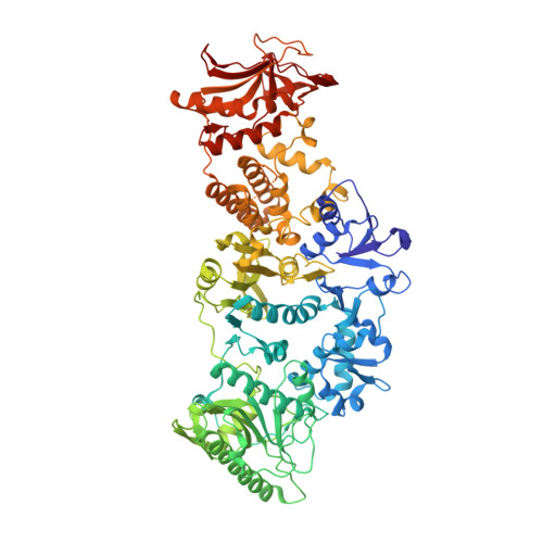
2.7 angstrom cryo-EM structure of rotavirus core protein VP3, a unique capping machine with a helicase activity.

Kumar, D.Yu, X.Crawford, S.E.Moreno, R.Jakana, J.Sankaran, B.Anish, R.Kaundal, S.Hu, L.Estes, M.K.Wang, Z.Prasad, B.V.V.

(2020) Sci Adv 6: eaay6410-eaay6410

  • DOI: https://doi.org/10.1126/sciadv.aay6410
  • Primary Citation of Related Structures:  
    6O3V

  • PubMed Abstract: 

    In many viruses, including rotavirus (RV), the major pathogen of infantile gastroenteritis, capping of viral messenger RNAs is a pivotal step for efficient translation of the viral genome. In RV, VP3 caps the nascent transcripts synthesized from the genomic dsRNA segments by the RV polymerase VP1 within the particle core. Here, from cryo-electron microscopy, x-ray crystallography, and biochemical analyses, we show that VP3 forms a stable tetrameric assembly with each subunit having a modular domain organization, which uniquely integrates five distinct enzymatic steps required for capping the transcripts. In addition to the previously known guanylyl- and methyltransferase activities, we show that VP3 exhibits hitherto unsuspected RNA triphosphatase activity necessary for initiating transcript capping and RNA helicase activity likely required for separating the RNA duplex formed transiently during endogenous transcription. From our studies, we propose a new mechanism for how VP3 inside the virion core caps the nascent transcripts exiting from the polymerase.


  • Organizational Affiliation

    Verna and Marrs McLean Department of Biochemistry and Molecular Biology, Baylor College of Medicine, Houston, TX 77030, USA.


Macromolecules
Find similar proteins by:  (by identity cutoff)  |  3D Structure
Entity ID: 1
MoleculeChains Sequence LengthOrganismDetailsImage
Protein VP3
A, B, C
835Rotavirus AMutation(s): 0 
Gene Names: VP3
UniProt
Find proteins for Q1WK45 (Rotavirus A)
Explore Q1WK45 
Go to UniProtKB:  Q1WK45
Entity Groups  
Sequence Clusters30% Identity50% Identity70% Identity90% Identity95% Identity100% Identity
UniProt GroupQ1WK45
Sequence Annotations
Expand
  • Reference Sequence
Small Molecules
Ligands 3 Unique
IDChains Name / Formula / InChI Key2D Diagram3D Interactions
5GP
Query on 5GP

Download Ideal Coordinates CCD File 
D [auth A],
G [auth B],
J [auth C]
GUANOSINE-5'-MONOPHOSPHATE
C10 H14 N5 O8 P
RQFCJASXJCIDSX-UUOKFMHZSA-N
SO4
Query on SO4

Download Ideal Coordinates CCD File 
F [auth A],
I [auth B],
L [auth C]
SULFATE ION
O4 S
QAOWNCQODCNURD-UHFFFAOYSA-L
EDO
Query on EDO

Download Ideal Coordinates CCD File 
E [auth A],
H [auth B],
K [auth C]
1,2-ETHANEDIOL
C2 H6 O2
LYCAIKOWRPUZTN-UHFFFAOYSA-N
Experimental Data & Validation

Experimental Data

  • Method: X-RAY DIFFRACTION
  • Resolution: 3.50 Å
  • R-Value Free: 0.264 
  • R-Value Work: 0.229 
  • R-Value Observed: 0.231 
  • Space Group: P 62 2 2
Unit Cell:
Length ( Å )Angle ( ˚ )
a = 236.706α = 90
b = 236.706β = 90
c = 349.3γ = 120
Software Package:
Software NamePurpose
PHENIXrefinement
HKL-2000data reduction
HKL-2000data scaling
PHASERphasing

Structure Validation

View Full Validation Report


Ligand Structure Quality Assessment 


Entry History & Funding Information

Deposition Data


Funding OrganizationLocationGrant Number
National Institutes of Health/National Institute Of Allergy and Infectious Diseases (NIH/NIAID)United StatesAI36040
Welch FoundationUnited StatesQ1279

Revision History  (Full details and data files)

  • Version 1.0: 2020-03-04
    Type: Initial release
  • Version 1.1: 2020-09-16
    Changes: Database references
  • Version 1.2: 2024-03-13
    Changes: Data collection, Database references