6NTD | pdb_00006ntd

Crystal Structure of G12V HRas-GppNHp bound in complex with the engineered RBD variant 12 of CRAF Kinase protein


Experimental Data Snapshot

  • Method: X-RAY DIFFRACTION
  • Resolution: 3.15 Å
  • R-Value Free: 
    0.287 (Depositor), 0.306 (DCC) 
  • R-Value Work: 
    0.273 (Depositor) 
  • R-Value Observed: 
    0.274 (Depositor) 

Starting Model: experimental
View more details

wwPDB Validation 3D Report Full Report

Validation slider image for 6NTD

Ligand Structure Quality Assessment 


This is version 1.2 of the entry. See complete history

Literature

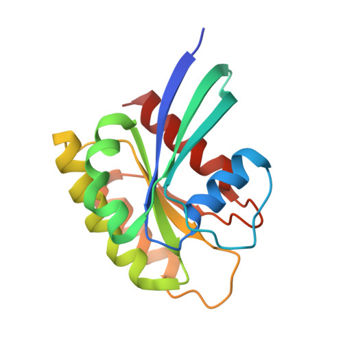
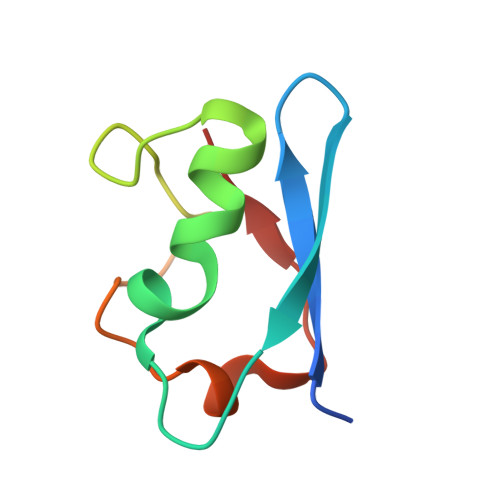
Conformation-specific inhibitors of activated Ras GTPases reveal limited Ras dependency of patient-derived cancer organoids.

Wiechmann, S.Maisonneuve, P.Grebbin, B.M.Hoffmeister, M.Kaulich, M.Clevers, H.Rajalingam, K.Kurinov, I.Farin, H.F.Sicheri, F.Ernst, A.

(2020) J Biological Chem 295: 4526-4540

  • DOI: https://doi.org/10.1074/jbc.RA119.011025
  • Primary Citation Related Structures: 
    6NTC, 6NTD

  • PubMed Abstract: 

    The small GTPases H, K, and NRAS are molecular switches indispensable for proper regulation of cellular proliferation and growth. Several mutations in the genes encoding members of this protein family are associated with cancer and result in aberrant activation of signaling processes caused by a deregulated recruitment of downstream effector proteins. In this study, we engineered variants of the Ras-binding domain (RBD) of the C-Raf proto-oncogene, Ser/Thr kinase (CRAF). These variants bound with high affinity with the effector-binding site of Ras in an active conformation. Structural characterization disclosed how the newly identified RBD mutations cooperate and thereby enhance affinity with the effector-binding site in Ras compared with WT RBD. The engineered RBD variants closely mimicked the interaction mode of naturally occurring Ras effectors and acted as dominant-negative affinity reagents that block Ras signal transduction. Experiments with cancer cells showed that expression of these RBD variants inhibits Ras signaling, reducing cell growth and inducing apoptosis. Using these optimized RBD variants, we stratified patient-derived colorectal cancer organoids with known Ras mutational status according to their response to Ras inhibition. These results revealed that the presence of Ras mutations was insufficient to predict sensitivity to Ras inhibition, suggesting that not all of these tumors required Ras signaling for proliferation. In summary, by engineering the Ras/Raf interface of the CRAF-RBD, we identified potent and selective inhibitors of Ras in its active conformation that outcompete binding of Ras-signaling effectors.


  • Organizational Affiliation
    • Institute of Biochemistry II, Goethe University Frankfurt-Medical Faculty, University Hospital, 60596 Frankfurt am Main, Germany.

Macromolecule Content 

  • Total Structure Weight: 29.08 kDa 
  • Atom Count: 1,665 
  • Modeled Residue Count: 231 
  • Deposited Residue Count: 252 
  • Unique protein chains: 2

Macromolecules

Find similar proteins by:|  3D Structure
Entity ID: 1
MoleculeChains  Sequence LengthOrganismDetailsImage
GTPase HRas171Homo sapiensMutation(s): 1 
Gene Names: HRASHRAS1
EC: 3.6.5.2
UniProt & NIH Common Fund Data Resources
Find proteins for P01112 (Homo sapiens)
Explore P01112 
Go to UniProtKB:  P01112
PHAROS:  P01112
GTEx:  ENSG00000174775 
Entity Groups
Sequence Clusters30% Identity50% Identity70% Identity90% Identity95% Identity100% Identity
UniProt GroupP01112
Sequence Annotations
Expand
Reference Sequence
Find similar proteins by:|  3D Structure
Entity ID: 2
MoleculeChains  Sequence LengthOrganismDetailsImage
RAF proto-oncogene serine/threonine-protein kinase81Homo sapiensMutation(s): 9 
Gene Names: RAF1RAF
EC: 2.7.11.1
UniProt & NIH Common Fund Data Resources
Find proteins for P04049 (Homo sapiens)
Explore P04049 
Go to UniProtKB:  P04049
PHAROS:  P04049
GTEx:  ENSG00000132155 
Entity Groups
Sequence Clusters30% Identity50% Identity70% Identity90% Identity95% Identity100% Identity
UniProt GroupP04049
Sequence Annotations
Expand
Reference Sequence

Small Molecules

Ligands 2 Unique
IDChains Name / Formula / InChI Key2D Diagram3D Interactions
GNP

Query on GNP


Download:Ideal Coordinates CCD File
D [auth A]PHOSPHOAMINOPHOSPHONIC ACID-GUANYLATE ESTER
C10 H17 N6 O13 P3
UQABYHGXWYXDTK-UUOKFMHZSA-N
MG

Query on MG


Download:Ideal Coordinates CCD File
C [auth A]MAGNESIUM ION
Mg
JLVVSXFLKOJNIY-UHFFFAOYSA-N

Experimental Data & Validation

Experimental Data

  • Method: X-RAY DIFFRACTION
  • Resolution: 3.15 Å
  • R-Value Free:  0.287 (Depositor), 0.306 (DCC) 
  • R-Value Work:  0.273 (Depositor) 
  • R-Value Observed: 0.274 (Depositor) 
Space Group: P 63 2 2
Unit Cell:
Length ( Å )Angle ( ˚ )
a = 91.761α = 90
b = 91.761β = 90
c = 151.59γ = 120
Software Package:
Software NamePurpose
PHENIXrefinement
XDSdata reduction
Aimlessdata scaling
PHASERphasing
Cootmodel building
PDB_EXTRACTdata extraction

Structure Validation

View Full Validation Report


Ligand Structure Quality Assessment 


Entry History 

Deposition Data

Revision History  (Full details and data files)

  • Version 1.0: 2020-03-04
    Type: Initial release
  • Version 1.1: 2020-04-15
    Changes: Database references
  • Version 1.2: 2023-10-11
    Changes: Data collection, Database references, Refinement description