6NMJ | pdb_00006nmj

Crystal Structure of Rat Ric-8A G alpha binding domain, "Paratone-N Immersed"


Experimental Data Snapshot

  • Method: X-RAY DIFFRACTION
  • Resolution: 2.30 Å
  • R-Value Free: 
    0.259 (Depositor), 0.263 (DCC) 
  • R-Value Work: 
    0.208 (Depositor), 0.211 (DCC) 
  • R-Value Observed: 
    0.209 (Depositor) 

Starting Model: experimental
View more details

wwPDB Validation 3D Report Full Report

Validation slider image for 6NMJ

This is version 1.3 of the entry. See complete history

Literature

Structure, Function, and Dynamics of the G alpha Binding Domain of Ric-8A.

Zeng, B.Mou, T.C.Doukov, T.I.Steiner, A.Yu, W.Papasergi-Scott, M.Tall, G.G.Hagn, F.Sprang, S.R.

(2019) Structure 27: 1137-1147.e5

  • DOI: https://doi.org/10.1016/j.str.2019.04.013
  • Primary Citation Related Structures: 
    6NMG, 6NMJ

  • PubMed Abstract: 

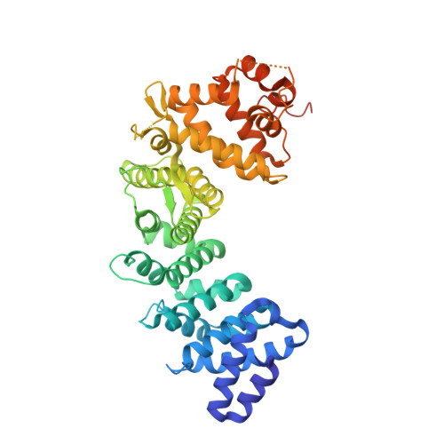
    Ric-8A is a 530-amino acid cytoplasmic molecular chaperone and guanine nucleotide exchange factor (GEF) for i, q, and 12/13 classes of heterortrimeric G protein alpha subunits (Gα). We report the 2.2-Å crystal structure of the Ric-8A Gα-binding domain with GEF activity, residues 1-452, and is phosphorylated at Ser435 and Thr440. Residues 1-429 adopt a superhelical fold comprised of Armadillo (ARM) and HEAT repeats, and the C terminus is disordered. One of the phosphorylated residues potentially binds to a basic cluster in an ARM motif. Amino acid sequence conservation and published hydrogen-deuterium exchange data indicate repeats 3 through 6 to be a putative Gα-binding surface. Normal mode modeling of small-angle X-ray scattering data indicates that phosphorylation induces relative rotation between repeats 1-4, 5-6, and 7-9. 2D 1 H- 15 N-TROSY spectra of [ 2 H, 15 N]-labeled Gαi1 in the presence of R452 reveals chemical shift perturbations of the C terminus and Gαi1 residues involved in nucleotide binding.


  • Organizational Affiliation
    • Graduate Program in Biochemistry and Biophysics, University of Montana, Missoula, MT 59812, USA.

Macromolecule Content 

  • Total Structure Weight: 102.32 kDa 
  • Atom Count: 6,541 
  • Modeled Residue Count: 813 
  • Deposited Residue Count: 906 
  • Unique protein chains: 1

Macromolecules

Find similar proteins by:|  3D Structure
Entity ID: 1
MoleculeChains  Sequence LengthOrganismDetailsImage
Resistance to inhibitors of cholinesterase 8 homolog A (C. elegans)
A, B
453Rattus norvegicusMutation(s): 1 
Gene Names: Ric8arCG_48458
UniProt
Find proteins for Q80ZG1 (Rattus norvegicus)
Explore Q80ZG1 
Go to UniProtKB:  Q80ZG1
Entity Groups
Sequence Clusters30% Identity50% Identity70% Identity90% Identity95% Identity100% Identity
UniProt GroupQ80ZG1
Sequence Annotations
Expand
Reference Sequence

Experimental Data & Validation

Experimental Data

  • Method: X-RAY DIFFRACTION
  • Resolution: 2.30 Å
  • R-Value Free:  0.259 (Depositor), 0.263 (DCC) 
  • R-Value Work:  0.208 (Depositor), 0.211 (DCC) 
  • R-Value Observed: 0.209 (Depositor) 
Space Group: P 21 21 21
Unit Cell:
Length ( Å )Angle ( ˚ )
a = 63.298α = 90
b = 100.114β = 90
c = 129.959γ = 90
Software Package:
Software NamePurpose
PHENIXrefinement
XDSdata reduction
XDSdata scaling
PHASERphasing

Structure Validation

View Full Validation Report


Entry History 

& Funding Information

Deposition Data


Funding OrganizationLocationGrant Number
National Institutes of Health/National Institute of General Medical Sciences (NIH/NIGMS)United States5R01GM105993
National Institutes of Health/National Institute of General Medical Sciences (NIH/NIGMS)United StatesP20GM103546

Revision History  (Full details and data files)

  • Version 1.0: 2019-06-26
    Type: Initial release
  • Version 1.1: 2019-09-25
    Changes: Data collection, Database references
  • Version 1.2: 2020-01-01
    Changes: Author supporting evidence
  • Version 1.3: 2023-10-11
    Changes: Data collection, Database references, Refinement description