6NCZ

Crystal structure of hybrid beta-glucuronidase/beta-galacturonidase from Fusicatenibacter saccharivorans bound to phenyl-thio-beta-D-glucuronide


Experimental Data Snapshot

  • Method: X-RAY DIFFRACTION
  • Resolution: 2.20 Å
  • R-Value Free: 0.218 
  • R-Value Work: 0.181 
  • R-Value Observed: 0.181 

wwPDB Validation   3D Report Full Report

Currently 6NCZ does not have a validation slider image.


This is version 2.1 of the entry. See complete history


Literature

Selecting a Single Stereocenter: The Molecular Nuances That Differentiate beta-Hexuronidases in the Human Gut Microbiome.

Pellock, S.J.Walton, W.G.Redinbo, M.R.

(2019) Biochemistry 58: 1311-1317

  • DOI: https://doi.org/10.1021/acs.biochem.8b01285
  • Primary Citation of Related Structures:  
    6NCW, 6NCX, 6NCY, 6NCZ

  • PubMed Abstract: 

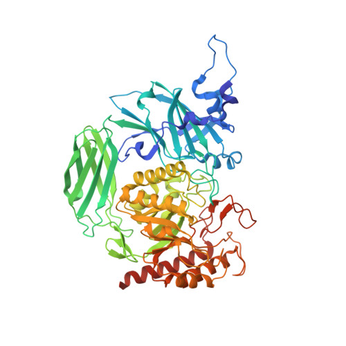
    The human gut microbiome is a ripe space for the discovery of new proteins and novel functions. Many genes in the gut microbiome encode glycoside hydrolases that help bacteria scavenge sugars present in the human gut. Glycoside hydrolase family 2 (GH2) is one group of sugar-scavenging proteins, which includes β-glucuronidases (GUS) and β-galacturonidases (GalAses), enzymes that cleave the sugar conjugates of the epimers glucuronate and galacturonate. Here we structurally and functionally characterize a GH2 GalAse and a hybrid GUS/GalAse, which reveal the molecular details that enable these GHs to differentiate a single stereocenter. First, we characterized a previously annotated GUS from Eisenbergiella tayi and demonstrated that it is, in fact, a GalAse. We determined the crystal structure of this GalAse, identified the key residue that confers GalAse activity, and convert this GalAse into a GUS by mutating a single residue. We performed bioinformatic analysis of 279 putative GUS enzymes from the human gut microbiome and identified 12 additional putative GH2 GalAses, one of which we characterized and confirmed is a GalAse. Lastly, we report the structure of a hybrid GUS/GalAse from Fusicatenibacter saccharivorans, which revealed a unique hexamer that positions the N-terminus of adjacent protomers in the aglycone binding site. Taken together, these data reveal a new class of bacterial GalAses in the human gut microbiome and unravel the structural details that differentiate GH2 GUSs and GalAses.


Macromolecules
Find similar proteins by:  (by identity cutoff)  |  3D Structure
Entity ID: 1
MoleculeChains Sequence LengthOrganismDetailsImage
Beta-glucuronidase
A, B, C, D, E
A, B, C, D, E, F
610Fusicatenibacter saccharivoransMutation(s): 0 
Gene Names: uidA_2ERS852406_01826
EC: 3.2.1.31
UniProt
Find proteins for A0A174EHD1 (Fusicatenibacter saccharivorans)
Explore A0A174EHD1 
Go to UniProtKB:  A0A174EHD1
Entity Groups  
Sequence Clusters30% Identity50% Identity70% Identity90% Identity95% Identity100% Identity
UniProt GroupA0A174EHD1
Sequence Annotations
Expand
  • Reference Sequence
Small Molecules
Ligands 2 Unique
IDChains Name / Formula / InChI Key2D Diagram3D Interactions
FYJ
Query on FYJ

Download Ideal Coordinates CCD File 
G [auth A]
I [auth B]
K [auth C]
M [auth D]
O [auth E]
G [auth A],
I [auth B],
K [auth C],
M [auth D],
O [auth E],
Q [auth F]
phenyl 1-thio-beta-D-glucopyranosiduronic acid
C12 H14 O6 S
XXJOOMMTHULGSD-LIJGXYGRSA-N
GOL
Query on GOL

Download Ideal Coordinates CCD File 
H [auth A]
J [auth B]
L [auth C]
N [auth D]
P [auth E]
H [auth A],
J [auth B],
L [auth C],
N [auth D],
P [auth E],
R [auth F]
GLYCEROL
C3 H8 O3
PEDCQBHIVMGVHV-UHFFFAOYSA-N
Experimental Data & Validation

Experimental Data

  • Method: X-RAY DIFFRACTION
  • Resolution: 2.20 Å
  • R-Value Free: 0.218 
  • R-Value Work: 0.181 
  • R-Value Observed: 0.181 
  • Space Group: P 1
Unit Cell:
Length ( Å )Angle ( ˚ )
a = 109.134α = 60.32
b = 116.916β = 62.53
c = 117.604γ = 87.09
Software Package:
Software NamePurpose
PHENIXrefinement
XDSdata reduction
Aimlessdata scaling
PHENIXphasing

Structure Validation

View Full Validation Report

Currently 6NCZ does not have a validation slider image.


Entry History & Funding Information

Deposition Data


Funding OrganizationLocationGrant Number
National Institutes of Health/National Cancer Institute (NIH/NCI)United States--

Revision History  (Full details and data files)

  • Version 1.0: 2019-02-20
    Type: Initial release
  • Version 1.1: 2019-03-13
    Changes: Data collection, Database references
  • Version 1.2: 2019-12-04
    Changes: Author supporting evidence
  • Version 2.0: 2020-07-29
    Type: Remediation
    Reason: Carbohydrate remediation
    Changes: Atomic model, Data collection, Derived calculations
  • Version 2.1: 2023-10-11
    Changes: Data collection, Database references, Refinement description, Structure summary