6MUJ

Formylglycine generating enzyme bound to copper


Experimental Data Snapshot

  • Method: X-RAY DIFFRACTION
  • Resolution: 2.25 Å
  • R-Value Free: 0.246 
  • R-Value Work: 0.202 
  • R-Value Observed: 0.203 

wwPDB Validation   3D Report Full Report

Currently 6MUJ does not have a validation slider image.


This is version 1.4 of the entry. See complete history


Literature

Formylglycine-generating enzyme binds substrate directly at a mononuclear Cu(I) center to initiate O2activation.

Appel, M.J.Meier, K.K.Lafrance-Vanasse, J.Lim, H.Tsai, C.L.Hedman, B.Hodgson, K.O.Tainer, J.A.Solomon, E.I.Bertozzi, C.R.

(2019) Proc Natl Acad Sci U S A 116: 5370-5375

  • DOI: https://doi.org/10.1073/pnas.1818274116
  • Primary Citation of Related Structures:  
    6MUJ

  • PubMed Abstract: 

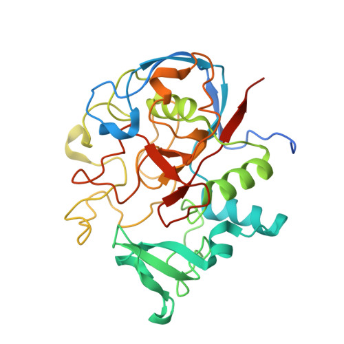
    The formylglycine-generating enzyme (FGE) is required for the posttranslational activation of type I sulfatases by oxidation of an active-site cysteine to C α -formylglycine. FGE has emerged as an enabling biotechnology tool due to the robust utility of the aldehyde product as a bioconjugation handle in recombinant proteins. Here, we show that Cu(I)-FGE is functional in O 2 activation and reveal a high-resolution X-ray crystal structure of FGE in complex with its catalytic copper cofactor. We establish that the copper atom is coordinated by two active-site cysteine residues in a nearly linear geometry, supporting and extending prior biochemical and structural data. The active cuprous FGE complex was interrogated directly by X-ray absorption spectroscopy. These data unambiguously establish the configuration of the resting enzyme metal center and, importantly, reveal the formation of a three-coordinate tris(thiolate) trigonal planar complex upon substrate binding as furthermore supported by density functional theory (DFT) calculations. Critically, inner-sphere substrate coordination turns on O 2 activation at the copper center. These collective results provide a detailed mechanistic framework for understanding why nature chose this structurally unique monocopper active site to catalyze oxidase chemistry for sulfatase activation.


  • Organizational Affiliation

    Department of Molecular and Cell Biology, University of California, Berkeley, CA 94720.


Macromolecules
Find similar proteins by:  (by identity cutoff)  |  3D Structure
Entity ID: 1
MoleculeChains Sequence LengthOrganismDetailsImage
Formylglycine-generating enzyme
A, B, C, D, E
319Streptomyces coelicolor A3(2)Mutation(s): 0 
Gene Names: SCO7548
EC: 1.8.3.7
UniProt
Find proteins for Q9F3C7 (Streptomyces coelicolor (strain ATCC BAA-471 / A3(2) / M145))
Explore Q9F3C7 
Go to UniProtKB:  Q9F3C7
Entity Groups  
Sequence Clusters30% Identity50% Identity70% Identity90% Identity95% Identity100% Identity
UniProt GroupQ9F3C7
Sequence Annotations
Expand
  • Reference Sequence
Small Molecules
Ligands 7 Unique
IDChains Name / Formula / InChI Key2D Diagram3D Interactions
DTT
Query on DTT

Download Ideal Coordinates CCD File 
HA [auth D]2,3-DIHYDROXY-1,4-DITHIOBUTANE
C4 H10 O2 S2
VHJLVAABSRFDPM-IMJSIDKUSA-N
GOL
Query on GOL

Download Ideal Coordinates CCD File 
AA [auth D],
Q [auth C],
X [auth C],
Y [auth C]
GLYCEROL
C3 H8 O3
PEDCQBHIVMGVHV-UHFFFAOYSA-N
GLY
Query on GLY

Download Ideal Coordinates CCD File 
Z [auth D]GLYCINE
C2 H5 N O2
DHMQDGOQFOQNFH-UHFFFAOYSA-N
IMD
Query on IMD

Download Ideal Coordinates CCD File 
IA [auth D],
K [auth A],
L [auth B],
OA [auth E]
IMIDAZOLE
C3 H5 N2
RAXXELZNTBOGNW-UHFFFAOYSA-O
CU
Query on CU

Download Ideal Coordinates CCD File 
CA [auth D]
DA [auth D]
EA [auth D]
FA [auth D]
G [auth A]
CA [auth D],
DA [auth D],
EA [auth D],
FA [auth D],
G [auth A],
GA [auth D],
H [auth A],
I [auth A],
J [auth A],
KA [auth E],
LA [auth E],
MA [auth E],
N [auth B],
NA [auth E],
O [auth B],
P [auth B],
S [auth C],
T [auth C],
U [auth C],
V [auth C]
COPPER (II) ION
Cu
JPVYNHNXODAKFH-UHFFFAOYSA-N
FMT
Query on FMT

Download Ideal Coordinates CCD File 
W [auth C]FORMIC ACID
C H2 O2
BDAGIHXWWSANSR-UHFFFAOYSA-N
CA
Query on CA

Download Ideal Coordinates CCD File 
BA [auth D],
F [auth A],
JA [auth E],
M [auth B],
R [auth C]
CALCIUM ION
Ca
BHPQYMZQTOCNFJ-UHFFFAOYSA-N
Experimental Data & Validation

Experimental Data

  • Method: X-RAY DIFFRACTION
  • Resolution: 2.25 Å
  • R-Value Free: 0.246 
  • R-Value Work: 0.202 
  • R-Value Observed: 0.203 
  • Space Group: P 31 2 1
Unit Cell:
Length ( Å )Angle ( ˚ )
a = 140.008α = 90
b = 140.008β = 90
c = 217.508γ = 120
Software Package:
Software NamePurpose
PHENIXrefinement
PDB_EXTRACTdata extraction
XDSdata reduction
Aimlessdata scaling
PHENIXphasing

Structure Validation

View Full Validation Report

Currently 6MUJ does not have a validation slider image.


Entry History & Funding Information

Deposition Data


Funding OrganizationLocationGrant Number
National Institutes of Health/National Cancer Institute (NIH/NCI)United StatesR35CA22043

Revision History  (Full details and data files)

  • Version 1.0: 2019-02-27
    Type: Initial release
  • Version 1.1: 2019-03-13
    Changes: Data collection, Database references
  • Version 1.2: 2019-03-27
    Changes: Data collection, Database references
  • Version 1.3: 2019-12-04
    Changes: Author supporting evidence
  • Version 1.4: 2023-10-11
    Changes: Data collection, Database references, Derived calculations, Refinement description