6MRC | pdb_00006mrc

ADP-bound human mitochondrial Hsp60-Hsp10 football complex


Experimental Data Snapshot

  • Method: ELECTRON MICROSCOPY
  • Resolution: 3.08 Å
  • Aggregation State: PARTICLE 
  • Reconstruction Method: SINGLE PARTICLE 

wwPDB Validation 3D Report Full Report

Validation slider image for 6MRC

This is version 1.4 of the entry. See complete history

Literature

Structural basis for active single and double ring complexes in human mitochondrial Hsp60-Hsp10 chaperonin.

Gomez-Llorente, Y.Jebara, F.Patra, M.Malik, R.Nisemblat, S.Chomsky-Hecht, O.Parnas, A.Azem, A.Hirsch, J.A.Ubarretxena-Belandia, I.

(2020) Nat Commun 11: 1916-1916

  • DOI: https://doi.org/10.1038/s41467-020-15698-8
  • Primary Citation Related Structures: 
    6MRC, 6MRD

  • PubMed Abstract: 

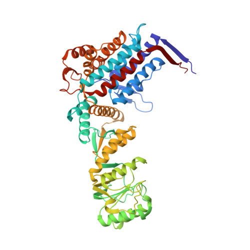
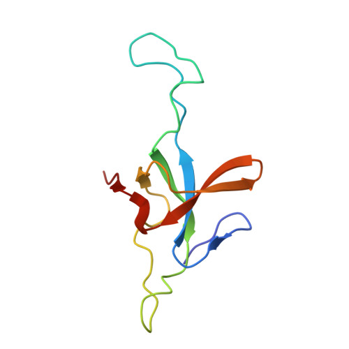
    mHsp60-mHsp10 assists the folding of mitochondrial matrix proteins without the negative ATP binding inter-ring cooperativity of GroEL-GroES. Here we report the crystal structure of an ATP (ADP:BeF 3 -bound) ground-state mimic double-ring mHsp60 14 -(mHsp10 7 ) 2 football complex, and the cryo-EM structures of the ADP-bound successor mHsp60 14 -(mHsp10 7 ) 2 complex, and a single-ring mHsp60 7 -mHsp10 7 half-football. The structures explain the nucleotide dependence of mHsp60 ring formation, and reveal an inter-ring nucleotide symmetry consistent with the absence of negative cooperativity. In the ground-state a two-fold symmetric H-bond and a salt bridge stitch the double-rings together, whereas only the H-bond remains as the equatorial gap increases in an ADP football poised to split into half-footballs. Refolding assays demonstrate obligate single- and double-ring mHsp60 variants are active, and complementation analysis in bacteria shows the single-ring variant is as efficient as wild-type mHsp60. Our work provides a structural basis for active single- and double-ring complexes coexisting in the mHsp60-mHsp10 chaperonin reaction cycle.


  • Organizational Affiliation
    • Department of Pharmacological Sciences, Icahn School of Medicine at Mount Sinai, New York, NY, 10029, USA.

Macromolecule Content 

  • Total Structure Weight: 944.43 kDa 
  • Atom Count: 66,178 
  • Modeled Residue Count: 8,792 
  • Deposited Residue Count: 8,792 
  • Unique protein chains: 2

Macromolecules

Find similar proteins by:|  3D Structure
Entity ID: 1
MoleculeChains  Sequence LengthOrganismDetailsImage
60 kDa heat shock protein, mitochondrial528Homo sapiensMutation(s): 0 
Gene Names: HSPD1HSP60
EC: 3.6.4.9 (PDB Primary Data), 5.6.1.7 (UniProt)
UniProt & NIH Common Fund Data Resources
Find proteins for P10809 (Homo sapiens)
Explore P10809 
Go to UniProtKB:  P10809
PHAROS:  P10809
GTEx:  ENSG00000144381 
Entity Groups
Sequence Clusters30% Identity50% Identity70% Identity90% Identity95% Identity100% Identity
UniProt GroupP10809
Sequence Annotations
Expand
Reference Sequence
Find similar proteins by:|  3D Structure
Entity ID: 2
MoleculeChains  Sequence LengthOrganismDetailsImage
10 kDa heat shock protein, mitochondrial100Homo sapiensMutation(s): 0 
Gene Names: HSPE1
UniProt & NIH Common Fund Data Resources
Find proteins for P61604 (Homo sapiens)
Explore P61604 
Go to UniProtKB:  P61604
PHAROS:  P61604
GTEx:  ENSG00000115541 
Entity Groups
Sequence Clusters30% Identity50% Identity70% Identity90% Identity95% Identity100% Identity
UniProt GroupP61604
Sequence Annotations
Expand
Reference Sequence

Small Molecules

Ligands 2 Unique
IDChains Name / Formula / InChI Key2D Diagram3D Interactions
ADP

Query on ADP


Download:Ideal Coordinates CCD File
AB [auth F]
CA [auth H]
CB [auth E]
EA [auth N]
GA [auth M]
AB [auth F],
CA [auth H],
CB [auth E],
EA [auth N],
GA [auth M],
IA [auth L],
KA [auth K],
MA [auth J],
OA [auth I],
QA [auth D],
SA [auth C],
UA [auth B],
WA [auth A],
YA [auth G]
ADENOSINE-5'-DIPHOSPHATE
C10 H15 N5 O10 P2
XTWYTFMLZFPYCI-KQYNXXCUSA-N
MG

Query on MG


Download:Ideal Coordinates CCD File
BB [auth F]
DA [auth H]
DB [auth E]
FA [auth N]
HA [auth M]
BB [auth F],
DA [auth H],
DB [auth E],
FA [auth N],
HA [auth M],
JA [auth L],
LA [auth K],
NA [auth J],
PA [auth I],
RA [auth D],
TA [auth C],
VA [auth B],
XA [auth A],
ZA [auth G]
MAGNESIUM ION
Mg
JLVVSXFLKOJNIY-UHFFFAOYSA-N

Experimental Data & Validation

Experimental Data

  • Method: ELECTRON MICROSCOPY
  • Resolution: 3.08 Å
  • Aggregation State: PARTICLE 
  • Reconstruction Method: SINGLE PARTICLE 
EM Software:
TaskSoftware PackageVersion
MODEL REFINEMENTPHENIX

Structure Validation

View Full Validation Report


Entry History 

& Funding Information

Deposition Data


Funding OrganizationLocationGrant Number
United States - Israel Binational Science Foundation (BSF)United States--

Revision History  (Full details and data files)

  • Version 1.0: 2020-04-15
    Type: Initial release
  • Version 1.1: 2020-05-06
    Changes: Database references
  • Version 1.2: 2020-07-08
    Changes: Structure summary
  • Version 1.3: 2024-03-13
    Changes: Data collection, Database references, Derived calculations
  • Version 1.4: 2025-05-14
    Changes: Data collection, Structure summary