6MNU

Crystal structure of Yersinia pestis UDP-glucose pyrophosphorylase


Experimental Data Snapshot

  • Method: X-RAY DIFFRACTION
  • Resolution: 2.17 Å
  • R-Value Free: 0.241 
  • R-Value Work: 0.177 
  • R-Value Observed: 0.179 

wwPDB Validation   3D Report Full Report

Currently 6MNU does not have a validation slider image.


This is version 1.1 of the entry. See complete history


Literature

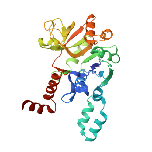
Crystal structure of UDP-glucose pyrophosphorylase from Yersinia pestis, a potential therapeutic target against plague.

Gibbs, M.E.Lountos, G.T.Gumpena, R.Waugh, D.S.

(2019) Acta Crystallogr F Struct Biol Commun 75: 608-615

  • DOI: https://doi.org/10.1107/S2053230X19011154
  • Primary Citation of Related Structures:  
    6MNU

  • PubMed Abstract: 

    Yersinia pestis, the causative agent of bubonic plague, is one of the most lethal pathogens in recorded human history. Today, the concern is the possible misuse of Y. pestis as an agent in bioweapons and bioterrorism. Current therapies for the treatment of plague include the use of a small number of antibiotics, but clinical cases of antibiotic resistance have been reported in some areas of the world. Therefore, the discovery of new drugs is required to combat potential Y. pestis infection. Here, the crystal structure of the Y. pestis UDP-glucose pyrophosphorylase (UGP), a metabolic enzyme implicated in the survival of Y. pestis in mouse macrophages, is described at 2.17 Å resolution. The structure provides a foundation that may enable the rational design of inhibitors and open new avenues for the development of antiplague therapeutics.


  • Organizational Affiliation

    Macromolecular Crystallography Laboratory, Center for Cancer Research, National Cancer Institute at Frederick, Frederick, MD 21702, USA.


Macromolecules
Find similar proteins by:  (by identity cutoff)  |  3D Structure
Entity ID: 1
MoleculeChains Sequence LengthOrganismDetailsImage
UTP--glucose-1-phosphate uridylyltransferase
A, B, C, D
296Yersinia pestisMutation(s): 0 
Gene Names: galUgalU1y2631YP_1428DBB30_10560EGT45_14485NCTC144_03080
EC: 2.7.7.9
UniProt
Find proteins for A0A3N4B738 (Yersinia pestis)
Explore A0A3N4B738 
Go to UniProtKB:  A0A3N4B738
Entity Groups  
Sequence Clusters30% Identity50% Identity70% Identity90% Identity95% Identity100% Identity
UniProt GroupA0A3N4B738
Sequence Annotations
Expand
  • Reference Sequence
Experimental Data & Validation

Experimental Data

  • Method: X-RAY DIFFRACTION
  • Resolution: 2.17 Å
  • R-Value Free: 0.241 
  • R-Value Work: 0.177 
  • R-Value Observed: 0.179 
  • Space Group: P 21 21 21
Unit Cell:
Length ( Å )Angle ( ˚ )
a = 92.19α = 90
b = 99.415β = 90
c = 128.656γ = 90
Software Package:
Software NamePurpose
PHENIXrefinement
HKL-3000data reduction
HKL-3000data scaling
PHASERphasing

Structure Validation

View Full Validation Report

Currently 6MNU does not have a validation slider image.


Entry History 

Deposition Data

Revision History  (Full details and data files)

  • Version 1.0: 2019-09-11
    Type: Initial release
  • Version 1.1: 2023-10-11
    Changes: Data collection, Database references, Refinement description