6ML4 | pdb_00006ml4

BTB24 Zinc Fingers 4-8 with 19+1mer DNA Oligonucleotide (Sequence 3)


Experimental Data Snapshot

  • Method: X-RAY DIFFRACTION
  • Resolution: 1.48 Å
  • R-Value Free: 
    0.211 (Depositor), 0.212 (DCC) 
  • R-Value Work: 
    0.191 (Depositor), 0.191 (DCC) 
  • R-Value Observed: 
    0.192 (Depositor) 

Starting Model: experimental
View more details

wwPDB Validation 3D Report Full Report

Validation slider image for 6ML4

This is version 1.3 of the entry. See complete history

Literature

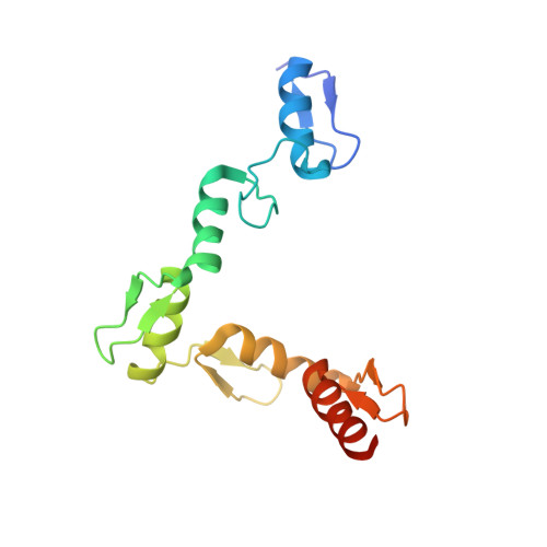
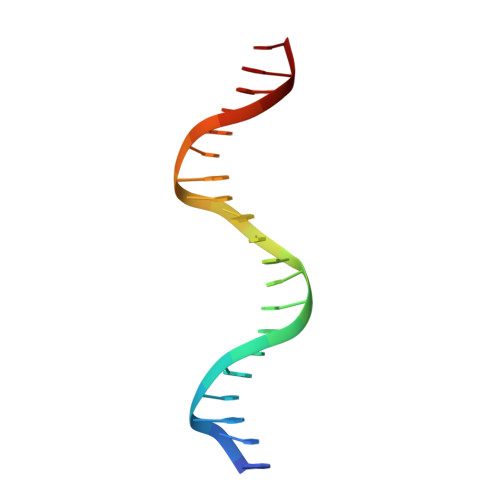
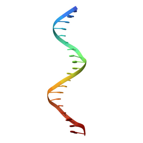
Structural basis of specific DNA binding by the transcription factor ZBTB24.

Ren, R.Hardikar, S.Horton, J.R.Lu, Y.Zeng, Y.Singh, A.K.Lin, K.Coletta, L.D.Shen, J.Lin Kong, C.S.Hashimoto, H.Zhang, X.Chen, T.Cheng, X.

(2019) Nucleic Acids Res 47: 8388-8398

  • DOI: https://doi.org/10.1093/nar/gkz557
  • Primary Citation Related Structures: 
    6ML2, 6ML3, 6ML4, 6ML5, 6ML6, 6ML7

  • PubMed Abstract: 

    ZBTB24, encoding a protein of the ZBTB family of transcriptional regulators, is one of four known genes-the other three being DNMT3B, CDCA7 and HELLS-that are mutated in immunodeficiency, centromeric instability and facial anomalies (ICF) syndrome, a genetic disorder characterized by DNA hypomethylation and antibody deficiency. The molecular mechanisms by which ZBTB24 regulates gene expression and the biological functions of ZBTB24 are poorly understood. Here, we identified a 12-bp consensus sequence [CT(G/T)CCAGGACCT] occupied by ZBTB24 in the mouse genome. The sequence is present at multiple loci, including the Cdca7 promoter region, and ZBTB24 binding is mostly associated with gene activation. Crystallography and DNA-binding data revealed that the last four of the eight zinc fingers (ZFs) (i.e. ZF5-8) in ZBTB24 confer specificity of DNA binding. Two ICF missense mutations have been identified in the ZBTB24 ZF domain, which alter zinc-binding cysteine residues. We demonstrated that the corresponding C382Y and C407G mutations in mouse ZBTB24 abolish specific DNA binding and fail to induce Cdca7 expression. Our analyses indicate and suggest a structural basis for the sequence specific recognition by a transcription factor centrally important for the pathogenesis of ICF syndrome.


  • Organizational Affiliation
    • Department of Epigenetics and Molecular Carcinogenesis, The University of Texas MD Anderson Cancer Center, Houston, TX 77030, USA.

Macromolecule Content 

  • Total Structure Weight: 30.06 kDa 
  • Atom Count: 2,337 
  • Modeled Residue Count: 183 
  • Deposited Residue Count: 191 
  • Unique protein chains: 1
  • Unique nucleic acid chains: 2

Macromolecules


Find similar proteins by:|  3D Structure
Entity ID: 1
MoleculeChains  Sequence LengthOrganismDetailsImage
Zinc finger and BTB domain-containing protein 24151Mus musculusMutation(s): 0 
Gene Names: Zbtb24Bif1Bsg1Znf450
UniProt & NIH Common Fund Data Resources
Find proteins for Q80X44 (Mus musculus)
Explore Q80X44 
Go to UniProtKB:  Q80X44
IMPC:  MGI:3039618
Entity Groups
Sequence Clusters30% Identity50% Identity70% Identity90% Identity95% Identity100% Identity
UniProt GroupQ80X44
Sequence Annotations
Expand
Reference Sequence
Find similar nucleic acids by:  Sequence
Entity ID: 2
MoleculeChains LengthOrganismImage
DNA (5'-D(*AP*CP*GP*CP*AP*GP*GP*TP*CP*CP*TP*GP*GP*AP*CP*GP*AP*AP*GP*C)-3')B [auth E]20Mus musculus
Sequence Annotations
Expand
Reference Sequence
Find similar nucleic acids by:  Sequence
Entity ID: 3
MoleculeChains LengthOrganismImage
DNA (5'-D(*TP*GP*CP*TP*TP*CP*GP*TP*CP*CP*AP*GP*GP*AP*CP*CP*TP*GP*CP*G)-3')C [auth F]20Mus musculus
Sequence Annotations
Expand
Reference Sequence

Small Molecules

Ligands 2 Unique
IDChains Name / Formula / InChI Key2D Diagram3D Interactions
ZN

Query on ZN


Download:Ideal Coordinates CCD File
D [auth A],
E [auth A],
F [auth A],
G [auth A],
H [auth A]
ZINC ION
Zn
PTFCDOFLOPIGGS-UHFFFAOYSA-N
EDO

Query on EDO


Download:Ideal Coordinates CCD File
I [auth A],
J [auth E],
K [auth E]
1,2-ETHANEDIOL
C2 H6 O2
LYCAIKOWRPUZTN-UHFFFAOYSA-N

Experimental Data & Validation

Experimental Data

  • Method: X-RAY DIFFRACTION
  • Resolution: 1.48 Å
  • R-Value Free:  0.211 (Depositor), 0.212 (DCC) 
  • R-Value Work:  0.191 (Depositor), 0.191 (DCC) 
  • R-Value Observed: 0.192 (Depositor) 
Space Group: P 1 21 1
Unit Cell:
Length ( Å )Angle ( ˚ )
a = 46.277α = 90
b = 68.807β = 92.4
c = 48.434γ = 90
Software Package:
Software NamePurpose
PHENIXrefinement
CrysalisProdata reduction
Aimlessdata scaling
PHENIXphasing

Structure Validation

View Full Validation Report


Entry History 

& Funding Information

Deposition Data


Funding OrganizationLocationGrant Number
National Institutes of Health/National Institute of General Medical Sciences (NIH/NIGMS)United StatesGM049245-23

Revision History  (Full details and data files)

  • Version 1.0: 2019-07-03
    Type: Initial release
  • Version 1.1: 2019-10-30
    Changes: Data collection, Database references
  • Version 1.2: 2020-01-01
    Changes: Author supporting evidence
  • Version 1.3: 2023-10-11
    Changes: Data collection, Database references, Refinement description