6MIM

Crystal structure of AF9 YEATS domain Y78W mutant in complex with histone H3K9cr


Experimental Data Snapshot

  • Method: X-RAY DIFFRACTION
  • Resolution: 2.52 Å
  • R-Value Free: 0.264 
  • R-Value Work: 0.203 
  • R-Value Observed: 0.209 

wwPDB Validation   3D Report Full Report


This is version 1.2 of the entry. See complete history


Literature

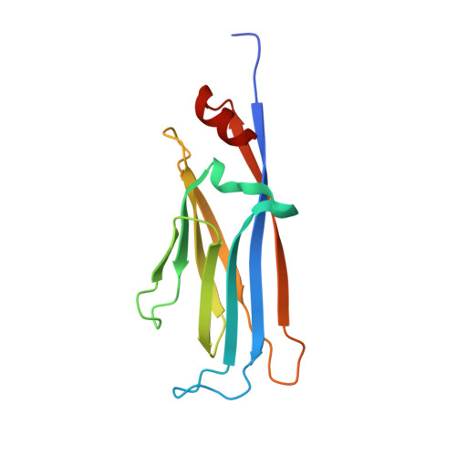
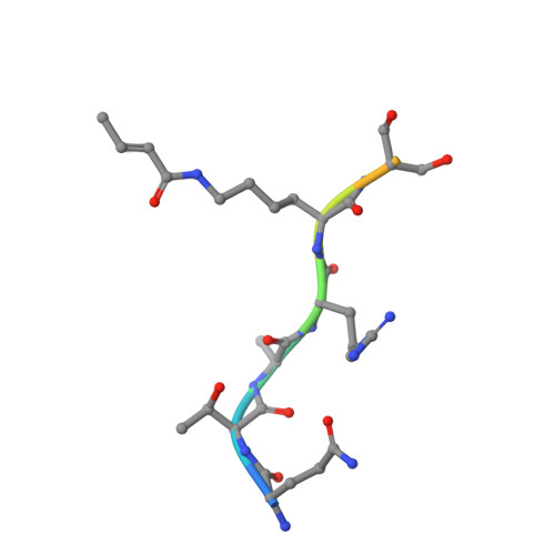
Structural insights into the pi-pi-pi stacking mechanism and DNA-binding activity of the YEATS domain.

Klein, B.J.Vann, K.R.Andrews, F.H.Wang, W.W.Zhang, J.Zhang, Y.Beloglazkina, A.A.Mi, W.Li, Y.Li, H.Shi, X.Kutateladze, A.G.Strahl, B.D.Liu, W.R.Kutateladze, T.G.

(2018) Nat Commun 9: 4574-4574

  • DOI: https://doi.org/10.1038/s41467-018-07072-6
  • Primary Citation of Related Structures:  
    6MIL, 6MIM, 6MIN, 6MIO, 6MIP, 6MIQ

  • PubMed Abstract: 

    The YEATS domain has been identified as a reader of histone acylation and more recently emerged as a promising anti-cancer therapeutic target. Here, we detail the structural mechanisms for π-π-π stacking involving the YEATS domains of yeast Taf14 and human AF9 and acylated histone H3 peptides and explore DNA-binding activities of these domains. Taf14-YEATS selects for crotonyllysine, forming π stacking with both the crotonyl amide and the alkene moiety, whereas AF9-YEATS exhibits comparable affinities to saturated and unsaturated acyllysines, engaging them through π stacking with the acyl amide. Importantly, AF9-YEATS is capable of binding to DNA, whereas Taf14-YEATS is not. Using a structure-guided approach, we engineered a mutant of Taf14-YEATS that engages crotonyllysine through the aromatic-aliphatic-aromatic π stacking and shows high selectivity for the crotonyl H3K9 modification. Our findings shed light on the molecular principles underlying recognition of acyllysine marks and reveal a previously unidentified DNA-binding activity of AF9-YEATS.


  • Organizational Affiliation

    Department of Pharmacology, University of Colorado School of Medicine, Aurora, CO, 80045, USA.


Macromolecules
Find similar proteins by:  (by identity cutoff)  |  3D Structure
Entity ID: 1
MoleculeChains Sequence LengthOrganismDetailsImage
Protein AF-9A,
B [auth C]
141Homo sapiensMutation(s): 1 
Gene Names: MLLT3AF9YEATS3
UniProt & NIH Common Fund Data Resources
Find proteins for P42568 (Homo sapiens)
Explore P42568 
Go to UniProtKB:  P42568
PHAROS:  P42568
GTEx:  ENSG00000171843 
Entity Groups  
Sequence Clusters30% Identity50% Identity70% Identity90% Identity95% Identity100% Identity
UniProt GroupP42568
Sequence Annotations
Expand
  • Reference Sequence

Find similar proteins by:  Sequence   |   3D Structure  

Entity ID: 2
MoleculeChains Sequence LengthOrganismDetailsImage
Histone H3K9crC [auth B],
D
10Homo sapiensMutation(s): 0 
UniProt & NIH Common Fund Data Resources
Find proteins for P68431 (Homo sapiens)
Explore P68431 
Go to UniProtKB:  P68431
PHAROS:  P68431
Entity Groups  
Sequence Clusters30% Identity50% Identity70% Identity90% Identity95% Identity100% Identity
UniProt GroupP68431
Sequence Annotations
Expand
  • Reference Sequence
Small Molecules
Modified Residues  1 Unique
IDChains TypeFormula2D DiagramParent
KCR
Query on KCR
C [auth B],
D
L-PEPTIDE LINKINGC10 H18 N2 O3LYS
Experimental Data & Validation

Experimental Data

  • Method: X-RAY DIFFRACTION
  • Resolution: 2.52 Å
  • R-Value Free: 0.264 
  • R-Value Work: 0.203 
  • R-Value Observed: 0.209 
  • Space Group: P 21 21 21
Unit Cell:
Length ( Å )Angle ( ˚ )
a = 42.159α = 90
b = 59.697β = 90
c = 155.395γ = 90
Software Package:
Software NamePurpose
PHENIXrefinement
HKL-3000data reduction
HKL-3000data scaling
PHASERphasing

Structure Validation

View Full Validation Report


Entry History 

Deposition Data

Revision History  (Full details and data files)

  • Version 1.0: 2018-11-14
    Type: Initial release
  • Version 1.1: 2023-10-11
    Changes: Data collection, Database references, Refinement description
  • Version 1.2: 2023-11-15
    Changes: Data collection